Introduction to periodontal microsurgery
Periodontal microsurgery is one of the many advances that have taken place in the field of surgical periodontal therapy. Microsurgery is described as a methodology, a modification, and refinement of existing surgical techniques that uses magnification to improve visualization and has implications for and applicability to all specialities1. Historically, the first microsurgery was performed by Carl Nylen, in an eye surgery using a binocular microscope in 1922 2. He is considered as the father of microsurgery. Since its introduction, microscope-assisted medicine has grown exponentially. At present, microsurgery is extensively used in all the fields of medicine.
Microsurgery was introduced into dentistry by Apotheker and Jako in 1978 and it was first ……. Contents available in the book ……. Contents available in the book ……. Contents available in the book ……. Contents available in the book ……. Contents available in the book ……

Periobasics: A Textbook of Periodontics and Implantology
The book is usually delivered within one week anywhere in India and within three weeks anywhere throughout the world.
India Users:
International Users:
Rationale for microsurgical techniques
There are many advantages of microsurgery over conventional surgeries in terms of precision, reduced trauma and post-operative patient comfort. The rationales in favor of microsurgery include,
- The conventional surgeries involve larger area including more soft and hard tissue manipulation which results in greater post-operative edema, inflammation, and pain. On the other hand, because of their small size, microsurgical instruments reduce the amount of trauma to the tissue, thus reducing post-operative edema, pain, and discomfort.
- Gentle handling of the tissue is an important factor in deciding the success of periodontal esthetic surgeries. More the tissue is manipulated more the healing is compromised. During the microsurgical procedure, there is minimal manipulation of the tissue which is especially advantageous in periodontal esthetic surgeries.
- The blood supply of the operated area is directly related to healing. Microscope guided surgeries allow avoiding certain incisions all together, which helps in better blood perfusion. For example, while harvesting connective tissue graft from the palate, magnification allows placement of the incision line parallel to the cervix of the teeth, avoiding a releasing incision that would deny flap from one source of its blood supply. Moreover, the healing is faster with minimal scar formation.
- While performing conventional surgical procedures, tissue is injured more by crushing than manipulation. The regular blunt instruments crush the graft while ……. Contents available in the book ……. Contents available in the book ……. Contents available in the book ……. Contents available in the book ……. Contents available in the book ……
- Less bleeding during microsurgery facilitates better vision and better control over the surgical procedure which ultimately results in the better surgical outcome and patient acceptance.
- Healing by primary intention is always preferred over healing by secondary intention. In microsurgical techniques, a perfect coaptation of the edges of the incision is achieved due to the magnification of the area. Using passive suturing with micro-sutures further, facilitate primary healing.
- The acceptance by the patients is greatly improved because of the minimal risk of scar formation, significantly reduced postoperative pain, edema, healing time, discomfort from the stitches and their reduced visibility.
Principles and elements of microsurgery
Most of the dental treatment rendered historically has been done with the naked eye or macroscopically. In microsurgery, the treatment is rendered under a microscope. It is aimed at improving the operator’s skills and to provide better results in operating those areas where precision and delicacy are required. As a treatment philosophy, it encompasses three principles 5,
1. Improvement of motor skills, thereby enhancing surgical ability.
2. An emphasis on passive wound closure with exact primary apposition of the wound edge.
3. The application of microsurgical instrumentation and suturing to reduce tissue trauma.
There are three primary elements of microsurgery,
1. Magnification.
2. Illumination.
3. Instruments.
These elements are also referred to as microsurgical triad 6. No microsurgical technique can be done if any of these three components is lacking. Magnification visual acuity is the ability to perceive two closely lying objects separately. It primarily depends on the density of cells packed on the retina, the electrophysiologic process of the image formation on the retina and illumination of the area operated.
Magnification helps in visualization of fine details of the area to be ……. Contents available in the book ……. Contents available in the book ……. Contents available in the book ……. Contents available in the book ……. Contents available in the book ……
Clinical philosophy:
One needs to practice and attain a level of experience with microsurgical techniques to competently perform various periodontal surgical procedures. A good microsurgery can achieve results which cannot be commonly achieved by regular surgeries. To adapt micro-surgery in regular practice one should be open to new ideas and techniques. One needs to practice precise tissue handling and wound closure suturing technique to master microsurgical techniques.
Hand control during microsurgery
It is important to understand the basic aspects of hand movement before we go into the details of microsurgical techniques. Primarily, long flexor and extensor muscles control the finger movement. The finger movement performed by these muscles is relatively crude but is markedly improved when the wrist is stabilized on a flat surface. The control over finger movements is further improved by placing the wrist in a dorsiflexion position angled approximately at 20 degrees. Muscle tremors are significantly reduced in this position 5, 7.
Physiological tremors:
Physiologic tremor is the uncontrolled movement arising from both the intended and unintended actions of our body. Awareness of this effect is further enhanced under magnification. These unwanted hand movements during microsurgery not only compromises the operator’s skill, but also jeopardizes the results of the surgery. There are many factors affecting physiological tremors, including anxiety, alcohol, smoking, recent exercise, hypoglycemia, caffeine, and medications. To avoid these tremors, microsurgeon should have a relaxed mind, comfortable posture, well-supported hand and stable hold on the instrument. The basic principles of patient and operator positioning as described in “Principles of instrumentation” should be followed. Along with following these principles, additional ……. Contents available in the book ……. Contents available in the book ……. Contents available in the book ……. Contents available in the book ……. Contents available in the book ……
Instrument grasp
The grasps used in microsurgery are the same as described in “Principles of instrumentation”. Primarily, pen grasp (also known as an internal precision grip) is used for microsurgeries because it provides more stability than any other grasp 8. The fingers make a tripod effect in this position which provides maximum stability to the instrument.
Microsurgical instruments
As the name indicates, microsurgery uses a microscopic magnification of the area to be operated. Along with this microsurgical instruments are used which cause minimum trauma and bleeding during the surgery.
Magnification systems
Presently, a wide range of simple and complex magnification systems are available for microsurgery. There are basically two types of magnification systems, loupes and surgical microscopes.
Loupes
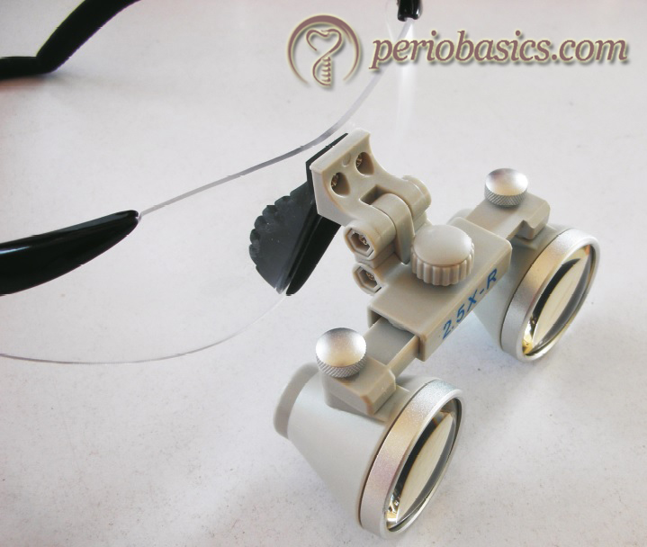
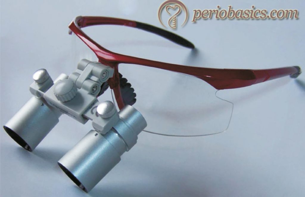
Dental loupes are the most common system used for optical magnification. These consist of two monocular microscopes with side-by-side lenses, angled to focus on one single object. A convergent lens optical system is called as Keplerian optical system. The major disadvantage associated with loupes is that eyes must converge to view an image which causes early eye straining and fatigue. Furthermore, prolonged use of loupes may result in vision change 9. Three types of loupes are commonly used,
1. Simple loupes
2. Compound loupes
3. Prism loupes
Simple loupes:
In these loupes, the magnification is achieved simply by adjustment of the working distance to a set length. It is referred to as diopter magnification. Simple loupes are primitive magnifiers with limited capabilities. The magnification can be increased by increasing the diameter and thickness of the lens. Because of size and weight ……. Contents available in the book ……. Contents available in the book ……. Contents available in the book ……. Contents available in the book ……. Contents available in the book ……
Compound Loupes:
These loupes use multiple lenses with intervening air space to gain additional refracting surfaces. These lenses can be adjusted to clinical needs without increasing their size or thickness. The lenses used in compound loupes can be achromatic which improves the optical design. In achromatic lenses, two pieces of lense are usually bonded together with a clear resin. The specific density of each piece counteracts the chromatic aberration of the adjacent piece. Clinicians should always choose compound loupes with achromatic lenses when selecting magnifying loupes. Magnification of compound loupes can be increased by lengthening the distance between the lenses. Compound loupes are commonly mounted on eyeglasses. A magnification up to 3X with these loupes provides a good quality image, beyond which the quality of the image is lost.
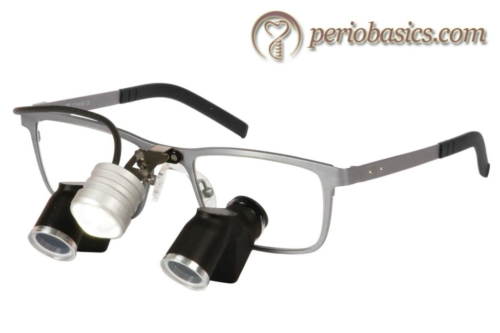
Prism loupes:
The prism loupes are most advanced loupe magnification systems. These contain Schmidt or rooftop prism which lengthens the light path through a series of switch back mirrors between the lenses. This arrangement folds the light so that the barrel of the loupes can be shortened. Furthermore, this arrangement produces better magnification, larger fields of view, wider depths of field, and longer working distances than do other loupes. Because of short barrel of the prism loupes, they can be mounted on ……. Contents available in the book ……. Contents available in the book ……. Contents available in the book ……. Contents available in the book ……. Contents available in the book ……
Advantages of loupes:
- These are less expensive.
- These are easy to use and are less likely to breach a clean operating field.
Limitations of loupes:
- These provide a limited magnification of the operating field, usually up to 4X. Beyond this magnification, the image gets distorted.
- Most of the loupes do not have an inbuilt light source, so an independent light source is required for illumination of the operative field.
- In loupes, there is approximately 4% loss of light because of surface refraction, unless anti-reflective coatings are placed on the lenses. In compound and prism loupes, the loss of brightness due to refraction may be up to 50% in the case where anti-reflective coatings are not placed.
- Loupes are inferior as compared to surgical microscopes when high visual acuity is required.
Surgical microscope
Surgical microscopes are superior to loupe microscopes in terms of flexibility, visual acuity, and magnification. However, these are much expensive than loupe magnifiers and are more difficult to use initially. The operating microscopes designed for use in dentistry are based on “Galilean principles”. The magnification provided by these micro-scopes ranges from 4X to 40X. The components of a surgical microscope are,
1. Magnification charger.
2. Objective lenses.
3. Binocular tubes.
4. Eyepieces.
5. Lightning unit.
6. Additional attachments.
Although surgical microscopes provide greatly improved visual acuity and the control of the surgical instruments, there are some basic problems associated with their use 12. While performing microsurgery, it is often required to visualize the defect from several angles to verify the debridement of areas of the osseous defect or the root surfaces. It is difficult ……. Contents available in the book ……. Contents available in the book ……. Contents available in the book ……. Contents available in the book ……. Contents available in the book ……
TOMS- Three-dimensional on-screen microsurgery system:
The current advances in the field of microsurgery allow visualization of the surgical site directly on the monitor. The clinician does not necessarily need to physically view through the microscope. The assembly of the TOMS consists of the surgical microscope, to which is attached a three-dimensional on-screen microsurgery system consisting of two single chip video cameras mounted on custom fit eyepiece adapters, a dual camera controller, a view/record image processor, a digital recording system, LCD/LED monitor for viewing the image of the surgical area, synchronizing signal emitter and 120 MHz shutter glasses (stereo eyewear) 13.
Surgical instruments
As a general rule, working end of all the instruments used during microsurgery should be smaller than the regular instruments. For high-quality performance during surgery, the average length of these instruments should be around 15 cm. The body of the instrument should be circular in cross section so that rotation while instrumentation is smooth. The working end or the cutting edge of these instruments should be smaller than that used for regular instruments. The instruments should preferably be made up of titanium, which makes them lighter and more resistant to distortion than regular stainless steel instruments.
In certain situations, instruments used in ophthalmic surgery are utilized. For example, while doing the papilla augmentation procedure, interdental papilla is dissected within the interdental embrasure. This incision is best placed by a “J” shaped ophthalmic blade, which is run under the interdental papilla and separates the tissue from the underlying bone. Once the incision is placed, the mini-flap can be raised by using a small periosteal elevator without tearing it. For stabilizing the tissue a small size ……. Contents available in the book ……. Contents available in the book ……. Contents available in the book ……. Contents available in the book ……. Contents available in the book ……
Micro-sutures
One of the basic rules in microsurgery is passive wound closure and exact primary apposition of the wound edges. For microsurgery, sutures with size 6-0 to 9-0 are used which are also referred to as micro-sutures. These sutures are primarily used in ophthalmic surgeries. The suture material is extremely thin and is difficult to visualize with naked eyes. Ideally, the size of the suture material and needle should be the same. After wound closure with precisely placed sutures, the incision line should be almost invisible to the naked eyes with minimal tissue damage and no bleeding.
Knot tying is an important part of the surgical procedure in microsurgeries. While placing a knot, the operator should be able to fully visualize his/her hands under macroscopic vision. The needle is held in the dominant hand and gloved fingertips of the other hand are used to place the tie using proprioception. The knot is placed by holding the suture with needle holder in the dominant hand and microsurgical tissue pick up in the non-dominant hand. While ……. Contents available in the book ……. Contents available in the book ……. Contents available in the book ……. Contents available in the book ……. Contents available in the book ……
Limitations of microsurgery (minimally invasive surgery)
Along with all the advantages of microsurgery discussed so far, there are some limitations also. Jaffray (2005) 14 enumerated following limitations of microsurgery,
1. It requires special equipment,
2. Specialized training is probably required,
3. Some additional equipment could be more expensive, and
4. Some procedures may take longer time than usual, as compared to the conventional surgeries.
Review of literature on microsurgery
Many studies have compared the outcome of the conventional surgeries and microsurgical procedures. In one such investigation, Cortellini and Tonetti (2001) 15 evaluated the outcomes of a microsurgical approach in the regenerative therapy of deep intrabony defects in 26 patients. They were treated with regenerative therapy using guided tissue regeneration. The results demonstrated that in 92.3% of the cases wound closure was intact throughout the healing period. The average clinical attachment gain was found to be 5.4 mm which ……. Contents available in the book ……. Contents available in the book ……. Contents available in the book ……. Contents available in the book ……. Contents available in the book ……
Another study was done by Wachtel et al. (2003) 16 to assess the clinical effect of the microsurgical access flap and enamel matrix derivatives (EMD) treatment with an emphasis on the evaluation of early wound healing. Patients included in the study had at least one pair of intrabony periodontal defects with an intrabony component of > or =3 mm. The parameters examined at baseline, 6 and 12 months after surgery included the oral hygiene status (API), gingival inflammation (BOP), probing pocket depth (PPD), clinical attachment level (CAL) and gingival recession (GR). The defects were randomly treated with ……. Contents available in the book ……. Contents available in the book ……. Contents available in the book ……. Contents available in the book ……. Contents available in the book ……
Harrel et al. (2005) 17 investigated the effect of using EMD in combination with minimally invasive surgery (MIS) in performing regenerative therapy in periodontal defects. The surgical treatment was performed in 160 sites on 16 patients. The results of the study demonstrated that the combination of MIS and EMD resulted in significant reductions in probing depths and improvements in attachment levels while producing little or no increase in recession.
A study done by Cortellini and Tonetti (2007) 18, evaluated clinical performance and patient perception associated with minimally invasive surgical technique in addition to the application of EMD in the treatment of isolated deep intra-bony defects. 13 deep intra-bony periodontal defects in 13 patients were surgically accessed with microsurgical technique. The authors concluded that in addition to a decrease in the surgical time, the technique decreased patient morbidity as the postoperative period was uneventful for 77% of the patients. In yet another study by Cortellini et al. (2008) 19, the clinical performance along with intra-operative and post-operative morbidity associated with minimally invasive surgical technique in addition to EMD application in the treatment of multiple deep intrabony defects in a single surgical procedure was evaluated. 44 defects in 20 patients were evaluated were surgically accessed. The results of the study suggested that MIS could be used in combination with EMD application in a single surgical procedure with excellent clinical outcomes with limited patient morbidity. A similar study was done by Jepsen et al. (2008) 20, where they evaluated ……. Contents available in the book ……. Contents available in the book ……. Contents available in the book ……. Contents available in the book ……. Contents available in the book ……
A modified minimally invasive surgical technique (M-MIST) was suggested by Cortellini and Tonetti (2009) 21 where only a buccal flap was raised while the interdental papilla was left in situ. The granulation tissue was removed, leaving the interdental and palatal tissues intact. After root instrumentation and placement of regenerative material, primary closure of the flap was achieved with a single internal modified mattress suture. The results of the study demonstrated limited patient morbidity and excellent clinical improvements. However, the authors suggested the requirement of a larger study to confirm the results as this study was done on 15 isolated interproximal defects out of 20 selected ones.
Conclusion
Microsurgical techniques have been shown to improve the treatment outcomes substantially and to a clinically relevant level as compared to conventional macrosurgical techniques. The magnification of the operative field allows the surgeon to view the area closely, facilitating precise and delicate tissue handling. However, the mastering of microsurgical techniques requires special training and practice. In addition, the cost of the equipment and instruments is an additional factor to be considered. New innovations are required in this field to make microsurgical techniques cost-effective for both the dentist and the patient.
References
References are available in the hard-copy of the website.
Periobasics: A Textbook of Periodontics and Implantology
The book is usually delivered within one week anywhere in India and within three weeks anywhere throughout the world.