Introduction to gingivectomy and gingivoplasty
The gingivectomy is the oldest surgical approach in periodontal therapy. During the centuries, the technique has been modified. At present, the technique of gingivectomy that is followed most commonly was given by Goldman HM (1951) 1. Gingivectomy means excision of the gingiva. It is a definitive surgical procedure indicated for pocket elimination in the presence of supra-bony pockets. Gingivoplasty, on the other hand, is the reshaping of the gingiva to create physiologic gingival contours with the sole purpose of recontouring the gingiva in the absence of pockets. Gingivectomy and gingivoplasty procedures are most commonly performed together. There are three prerequisites that must exist before gingivectomy is performed 2:
- The zone of gingiva must be wide enough so that excision of a part of it will still leave a functionally adequate zone,
- The underlying bone must be normal or near normal. If the bone loss has occurred, it must be horizontal in nature, leaving a relatively regular crestal bone at a lower level,
- There should not be infrabony defects or pockets.
Indications
Gingivectomy/gingivoplasty is indicated in the following conditions,
- Elimination of suprabony fibrous pockets and pseudo-pockets.
- Re-establishment of the physiological contour of gingiva.
- Elimination of fibrous or edematous enlargements of the gingiva.
- For improving the esthetics in cases where the anatomical crown has not been fully exposed.
- To create …….. Contents available in the book …….. Contents available in the book …….. Contents available in the book …….. Contents available in the book ……….
Contraindications
Gingivectomy is contraindicated in the following situations,
- In cases where the bottom of the pocket is apical to the mucogingival junction. In such cases, gingivectomy will result in an inadequate zone of attached gingiva.
- In areas with esthetic concern (maxillary and mandibular anterior areas) where gingivectomy may result in excessively long clinical crowns.
- Another relative contraindication is …….. Contents available in the book …….. Contents available in the book …….. Contents available in the book …….. Contents available in the book ……….
Periobasics: A Textbook of Periodontics and Implantology
The book is usually delivered within one week anywhere in India and within three weeks anywhere throughout the world.
India Users:
International Users:
Instruments
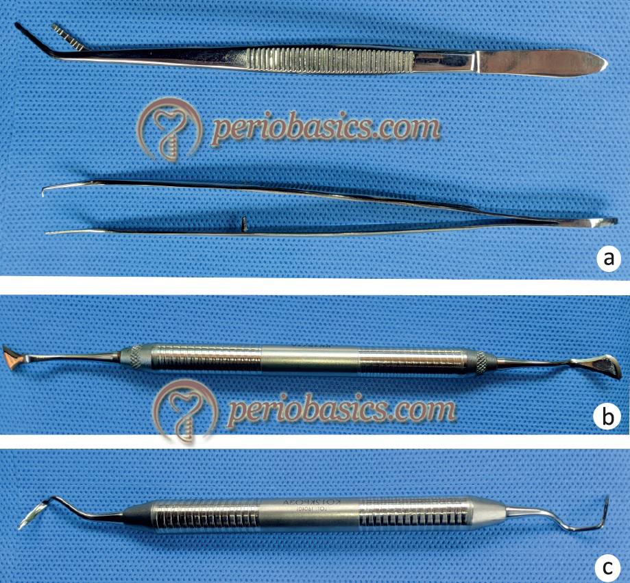
The surgical instruments used while performing gingivectomy include,
Pocket markers: Crane-Kaplan pocket marker/ Goldman-fox pocket marker.
Broad-bladed, round scalpels: Goldman fox No. 7/ Kirk-land knife.
Interproximal knife: Goldman fox No. 8, 9 & 10/ Orban’s knife.
Surgical handles: Bard parker handle no. 3 or angulated handle (Blake’s handle) with blade no. 11, 12, 15.
Curettes.
Tissue nippers.
These instruments are required for the traditional gingivectomy; however, gingivectomy can also be performed using round diamond burs, electrosurgery, and laser. In these procedures, the instruments used have been discussed with the respective surgical procedure.

Surgical technique
Gingivectomy can be performed by various methods. These methods include using gingivectomy knives or scalpel blade, electrosurgery or laser.
Gingivectomy using knives/blades:
A careful examination of the soft tissue and estimation of the probing depth is essential to visualize three-dimensional picture of the tissue which will facilitate a precise execution of the surgical procedure. Gingivectomy is performed in the following steps,
Marking the bleeding points:
The first step in gingivectomy is to mark the bleeding points once the area to be operated has been anesthetized. It is done with the help of a pocket marker (Crane-Kaplan / Goldman-fox pocket marker) (Figure 61.2). The pocket marker should be kept parallel to the long axis of the tooth. The beak of the pocket marker is inserted in the pocket up to the pocket depth and pinpoint perforations are created at the base of the pocket. These perforations …….. Contents available in the book …….. Contents available in the book …….. Contents available in the book …….. Contents available in the book ……….
Making incision:
Two types of incisions can be made during gingivectomy: Continuous and discontinuous. In both cases, the incision is made apical to the bottom of the bleeding point, starting at the most terminal tooth. An external bevel incision is made which is directed at 45ᵒ along the long axis of the tooth, coronally. The incision can be placed with Kirkland knife or BP blade No. 11/15 or Blake’s handle. Exactly how far apically from the bleeding points incision should be placed depends on the thickness of the gingiva (thicker the gingiva, more apical is the incision) and axial inclination of the teeth. In thick gingiva, a long bevel is produced; whereas, in thin gingiva, a small bevel is produced to achieve an appropriate scalloping form. The incision should be made in such a way that …….. Contents available in the book …….. Contents available in the book …….. Contents available in the book …….. Contents available in the book ……….


Removal of the excised tissue:
The excised tissue should be carefully removed with the help of a curette. If tissue tags are present, they should be removed with the help of scissors. If further reshaping of the tissue is required, it should be done with the help of rotating diamond burs.
Scaling and root planing:
Once the tissue has been removed, additional scaling and root planing should be done to remove any remaining calculus or necrotic cementum.
Placing periodontal dressing:
The periodontal dressing should be placed over the operated area after controlling the bleeding. This dressing should be removed one week after the surgical procedure. The patient should be given post-operative instructions and appropriate medications to control infection and pain should be prescribed to the patient.


Limiting factors associated with gingivectomy
It may be difficult to perform gingivectomy in the following situations,
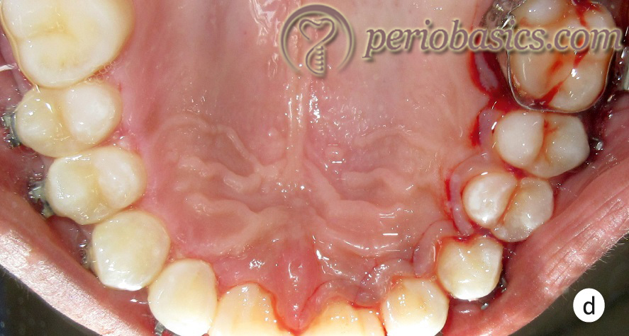
Palatal aspect of the maxillary posterior teeth:
In cases where the vault of the palate is shallow, the base of the periodontal pockets may be close to the vault area. In such cases, gingivectomy may result in excessive removal of the tissue. Hence, in such cases, the direction of the incision should be carefully planned.
Maxillary tuberosity area:
Performing gingivectomy in the maxillary tuberosity area to eliminate periodontal pockets is difficult because the tissue is abundant in this area. Furthermore, because of the limited access, it is difficult to place the external bevel incision in this area. In the maxillary tuberosity area, distal wedge procedure or distal molar surgical procedure is more appropriate.
Mandibular retromolar areas:
Similar to the maxillary tuberosity area, many times, the access to the mandibular retromolar area is limited. Furthermore, the tissue in this area is movable which makes it difficult to place the incision. Thus, the distal wedge procedure or distal molar surgical procedure is more appropriate in these areas.
Gingivectomy by electrosurgery
Gingivectomy can also be performed with electrosurgery. We require a needle electrode (thickness 0.0075 – 0.015 inch) and a small ovoid loop/diamond-shaped electrode to carry out the procedure. The area to be operated is anesthetized by administering a local anesthetic agent. The area is then isolated and dried a bit with a cotton swab. It must be noted here that if the area is too moist, considerable surface coagulation will occur; whereas, if the area is too dry excessive …….. Contents available in the book …….. Contents available in the book …….. Contents available in the book …….. Contents available in the book ……….
In cases where gingivectomy is done to eliminate excessive fibrotic gingival enlargement (such as drug-induced gingival enlargement), small ovoid loop electrodes are used to achieve proper festooning of the gingival margins. Most appropriate waveform suitable for gingivectomy is a fully rectified waveform. Once the procedure has been completed, the area should be cleaned with a moist gauge to remove all debris and periodontal dressing should be placed. The post-operative instructions are similar to those discussed previously. It should be noted here that electrosurgery should not be used …….. Contents available in the book …….. Contents available in the book …….. Contents available in the book …….. Contents available in the book ……….
Advantages:
The main advantage of electrosurgery is that the surgical procedure involves minimal or no bleeding. The incision is more precise as it is placed with no pressure. There is a minor loss of tissue after healing. These is minimal scar formation as the healing takes place by primary intention. The patient is more comfortable with this procedure as compared to conventional surgery with blades and knives.
Disadvantages:
As the procedure involves heat, there is generation of unpleasant odor. There are chances of damage to the bone or cementum, if the active electrode accidentally comes in contact with these tissues.
Gingivectomy using LASER
As discussed in, “Lasers in periodontice”, there are four main types of laser that are used in dentistry and they are different in the wavelengths of the emitted light energy. These types are: the Carbon dioxide laser (CO2) the Diode laser, the Neodymium: Aluminum-Yttrium-Garnet (Nd: YAG) and the Erbium: Aluminum-Yttrium-Garnet (Er: YAG) 3. All laser wave-lengths can be used to precisely incise gingiva; however, diode laser is most commonly used to perform gingivectomy.
Advantages:
Similar to electrosurgery, laser-assisted surgery is associated with minimal or no bleeding. The patient comfort and cooperation are very good with laser application. The incision placement is precise. Precise gingival contour can be achieved with laser application.
Disadvantages:
Laser-assisted gingivectomy is also associated with generation of unpleasant odor. Hence, high power vacuum suction is required during the procedure.
Healing after gingivectomy
After the tissue is excised, the operated area is covered by a blood clot. An acute inflammatory response is generated in the operated area which is characterized bya dilation of the blood vessels in the deeper tissue and migration of the leukocytes 4. Two days after the surgery, three layers can be observed in the blood clot 2: the necrotic surface layer, fibrinous innermost layer and leukocyte rich layer in between the outer and the inner layer. The epithelium starts growing under the clot at a rate of 0.5 mm/day, approximately 4 days after the surgery. In approximately two weeks, the entire surface is covered by epithelium.
During the first few days of healing, the connective tissue proliferation surrounding the blood vessels starts which is characterized by the mitotic activity in the fibroblasts, undifferentiated mesenchymal cells and endothelial cells 5. The proliferation of the connective tissue results in the formation of granulation tissue which then matures and the area subsequently gets …….. Contents available in the book …….. Contents available in the book …….. Contents available in the book …….. Contents available in the book ……….
Gingivoplasty
As already stated, the aim of gingivoplasty procedure is to recontour the gingiva that has lost its natural physiologic form. In contrast to gingivectomy that aims at the elimination of the supra-bony pockets, gingivoplasty aims at achieving the knife-edge gingival margins.
Gingivoplasty is indicated in the following situations,
- In cases with grossly thick gingival margins where normal gingival contour needs to be achieved.
- In cases with necrotizing ulcerative gingivitis/periodontitis, where after healing gingival clefts and craters are observed.
- In the presence of saucer-shaped deformities in the inter-dental areas of posterior teeth.
- Gingivoplasty can be performed with periodontal knives, diamond burs, electrosurgery or laser. The steps involved in gingivoplasty aim at thinning the gingival tissue to achieve a tapering gingival margin.
- It must be noted here that clinically, most of the times, gingivectomy and gingivoplasty are performed together.
Conclusion
Gingivectomy is the oldest surgical procedure in the field of periodontics. It is also one of the simplest surgical procedures. However, the incision should be placed carefully during gingivectomy because a failure to place a beveled incision results in the formation of a broad plateau because of which more time is required than usual to achieve the normal gingival contour. In the upcoming chapter, we shall read about various surgical techniques to treat periodontal pockets.
References
References are available in the hard-copy of the website.
Periobasics: A Textbook of Periodontics and Implantology
The book is usually delivered within one week anywhere in India and within three weeks anywhere throughout the world.

