Introduction to Bone morphogenetic proteins (BMPs):
Bone morphogenetic proteins (BMPs) are composed of 400-525 amino acids. These comprise one subset of the transforming growth factor-β superfamily. They play important roles in a multitude of processes during embryonic development and adult homeostasis by regulating cellular lineage commitment, morphogenesis, differentiation, proliferation, and apoptosis of various types of cells throughout the body. BMPs stimulate proliferation and migration of undifferentiated bone cell precursors with little to no effect on mature osteoprogenitor cells 1. Thus, the main action of BMPs is to commit ………. Contents available in the book ………. Contents available in the book ………. Contents available in the book ………. Contents available in the book ………

Periobasics: A Textbook of Periodontics and Implantology
The book is usually delivered within one week anywhere in India and within three weeks anywhere throughout the world.
India Users:
International Users:
Properties of bone morphogenetic proteins
- They act as mitogens on undifferentiated mesenchymal cells and osteoblast precursors.
- Structurally, they are related members of TGF-β super-family.
- BMP 2-12 singly initiate de novo endochondral bone formation 7-10.
- BMPs induce bone formation, whereas other growth factors such as TGF-β1 or PDGF do not.
- BMPs have an anabolic effect on periodontal tissue through the stimulation of osteoblastic differentiation in human periodontal ligament (PDL) cells 11.
- Bone allograft materials contain a varying amount of BMPs, such as BMP- 2,-4 and -7 12. The deficiency of BMP-like proteins retards bone cell differentiation and may account for a failure of the fracture to heal 13.
- Recombinant BMPs (rh BMPs) have been shown to promote bone formation 14, 15.
- They induce the expression of the osteoblast phenotype (i.e. increase in alkaline phosphatase activity in bone cells).
- Act as chemoattractants for mesenchymal cells and monocytes as well as bind to extracellular matrix collagen Type IV 16.
Historical Background
In 1889 Senn first found that decalcified ox bone promoted healing of osteomyelitic defects. He proposed that there was something in decalcified bone that was responsible for bone formation. In 1938 Lavender 17 implanted living bone fragments 1-1.5 cm in length, either subcutaneously or intramuscularly. These fragments were first treated by scraping away the periosteum, and in some specimens, a superficial layer of bone was also removed. Upon obtaining regenerated bone, he was able to show that it was neither the periosteum nor the cells on the surface or within the graft that were responsible for ………. Contents available in the book ………. Contents available in the book ………. Contents available in the book ………. Contents available in the book ………
In a series of experiments designed to test the triphasic theory of calcification, Urist et al. (1965, 1967) 7, 18 discovered that control samples of untreated, decalcified bone implanted into muscle pouches of rabbits and rats, resulted in new cartilage and bone formation by autoinduction. The purification of BMPs was completed using the rat ectopic bone formation assay. This assay involves combining the sample containing the unknown protein to be assayed with demineralized rat bone matrix, which has been treated with dissociative agents such as guanidine and urea to remove all of the endogenous BMP activity. This combination is then implanted subcutaneously in rats, and after 1-2 weeks, formation of new cartilage and bone is detected histologically. Using this bioassay, BMPs were purified and sequenced. Johnson et al. (1992) 19 successfully ………. Contents available in the book ………. Contents available in the book ………. Contents available in the book ………. Contents available in the book ………
Recombinant human BMP-2 through BMP- 6 and osteogenic protein-1 and -2 (OP-1 and OP-2, also known as BMP-7 and BMP-8, respectively), in conjunction with the collagenous matrix, singly induce de novo bone formation when implanted into extraskeletal sites of a variety of animal models 3, 22, 23. These recombinant BMPs have been produced using both mammalian cell expression system and Escherichia coli expression system.
Structure of BMPs
The BMPs are 30- to 38-kDa homodimers, glycosylated proteins. The individual BMP proteins are synthesized by a cell which then dimerize and become glycosylated. They are synthesized as prepropeptides of approximately 400-525 amino acids. The mature C-terminal region of 100-140 amino acid residues are released from a propeptide region by cleavage at an Arg-X-X-Arg sequence. Cleavage event occurs upon secretion that results in the formation of mature active protein, a dimer of the carboxy-terminal region of the precursor peptide. Because of the dimeric nature of the BMPs, it is possible that both homodimeric and heterodimeric forms exist. The BMPs have been grouped into subsets based on amino acid sequence homology. The groupings are suggested to be as follows:
(1) BMP-2 and BMP-4,
(2) BMP-3 and BMP-3b,
(3) BMP-5, BMP-6, BMP-7, and BMP-8,
(4) BMP-9 and BMP-10,
(5) BMP-12, BMP-13, and BMP-14, and
(6) BMP-11 and growth/differentiation factor 8 (GDF-8).
BMP signaling system
The BMPs signal through serine/threonine kinase receptors that are composed of type I and type II subtypes. Three type I receptors have been shown to bind ………. Contents available in the book ………. Contents available in the book ………. Contents available in the book ………. Contents available in the book ………
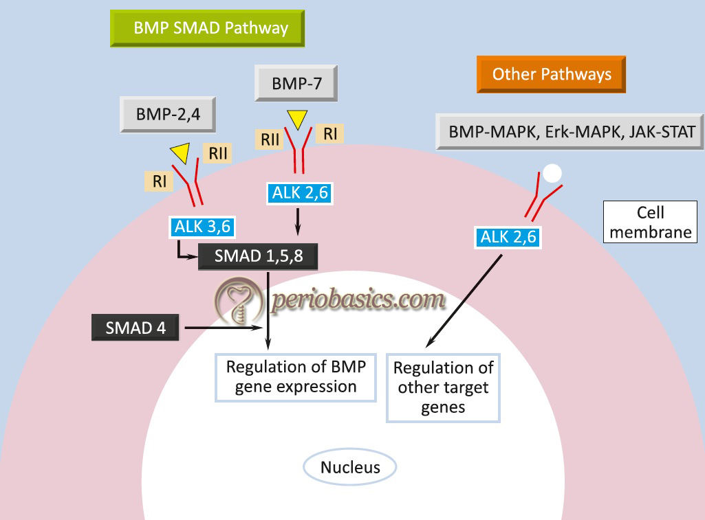
The type I BMP receptor substrates include the Smad proteins, which play a central role in the relay of BMP signals from the receptor to target genes in the nucleus. Smad1, Smad5, and Smad8 are phosphorylated by BMP receptors in a ligand-dependent manner. After release from the receptor, Smad proteins associate with the related protein Smad4, which acts as a shared partner. This complex translocates to the nucleus and participates in gene transcription with other transcription factors. Various BMPs perform different functions in body. Here is a brief description of some well-investigated BMPs,
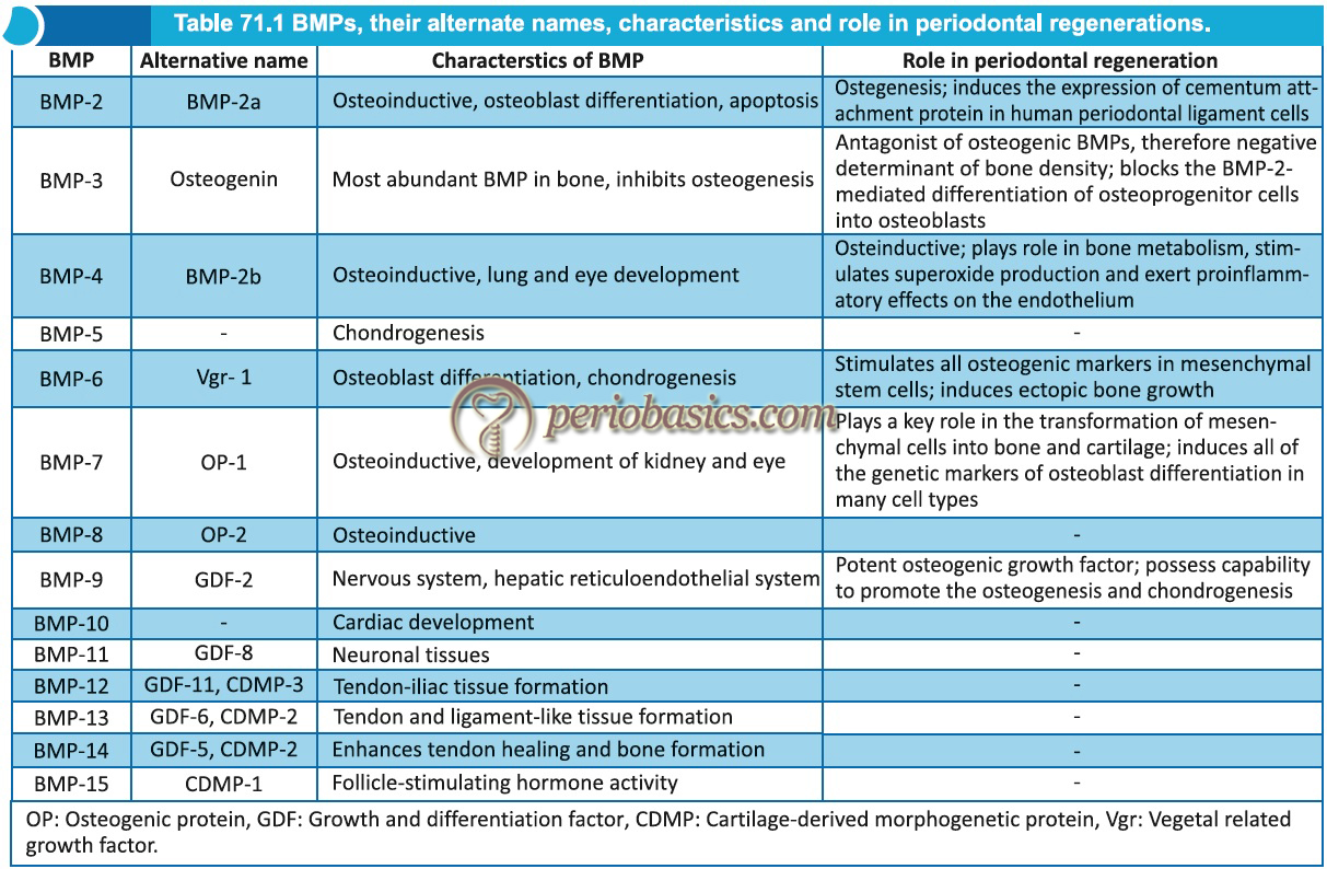
BMP-1
Genes for BMP 1 are located on chromosome 8. It does not belong to the TGF-β family of proteins. It is a metalloprotenase that acts on procollagen I, II, and III. It is involved in cartilage development.
BMP-2
It is a major inducer of osteoblast differentiation. The genes are located on chromosome 20.
BMP-3
Induces bone formation. The genes are located on chromosome 14.
BMP-4
Regulates the formation of teeth, limbs, and bone from mesoderm. It also plays a role in fracture repair. Genes are located on chromosome 14.
BMP-5
Performs functions in cartilage development. Genes are located on chromosome 6.
BMP-6
Plays a role in joint integrity in adults. Genes are located on chromosome 6.
BMP-7
Plays a key role in osteoblast differentiation. It also induces the production of Smad1. It is a key factor in renal development and repair. Genes are located on chromosome 20. It is also known as osteogenic protein-1 (OP-1).
Regulation of BMP signaling
The BMP signaling is regulated at multiple levels, both extracellularly and intracellularly. In extracellular compartments, BMP signaling is limited by BMP antagonists, which function through direct binding to BMP, thus preventing their binding to specific receptors. Similarly, there are various extracellular antagonists, which inhibit BMP activity. These include noggin, chordin, chordin-like 1, chordin-like 2, Gremlin, Cerberus, follistatin, and ectodin/ uterine sensitization-associated gene-1 (USAG-1). A negative feedback mechanism has been suggested wherein synthesis of antagonists such as noggin and Gremlin is up-regulated by BMPs. BMP signaling is also negatively regulated at the cell membrane by BAMBI ………. Contents available in the book ………. Contents available in the book ………. Contents available in the book ………. Contents available in the book ………
Role of BMPs in periodontal regeneration
BMPs have been used extensively by researchers to induce periodontal tissue regeneration in a variety of animal models 2, 24-27 as well as in human studies 28, 29 with varying degrees of success. Researchers 30 have proposed that BMPs possess a structure/activity profile with BMP-2 exhibiting mainly osteogenic properties while BMP-7 exhibiting mainly cementogenic activities.
Recombinant human bone morphogenetic protein-2 (rhBMP-2) has been used to investigate periodontal regeneration. Sigurdsson et al. (1995) 24 and Kinoshita et al. (1997) 27 successfully achieved periodontal regeneration in dogs using rhBMP-2 and a synthetic carrier. Clinical trials using ………. Contents available in the book ………. Contents available in the book ………. Contents available in the book ………. Contents available in the book ………
Challenges in clinical application of BMPs
A major challenge in the clinical application of BMPs is maintaining their optimum levels in the operated area for a sufficient duration of time. Furthermore, the activity of BMPs is non-specific and it has variable affects on different cell lineages in time and space 31, 32. The challenge in the clinical application of BMPs is the formulation of appropriate delivery systems for BMPs which is suitable for periodontal regeneration. The operational reconstitution and the implantation of the BMPs with the collagenous matrix as carrier, resulted in the induction of cementogenesis and morphogenesis of a functionally ………. Contents available in the book ………. Contents available in the book ………. Contents available in the book ………. Contents available in the book ………
Delivery systems for BMPs
As discussed in the previous section, there are many challenges in the clinical application of BMPs. There are primarily three approaches which have been used for the delivery of BMPs. These include gene-based, cell-based, and protein-based approach for BMP delivery (discussed in detail in “Tissue engineering in periodontics”). The gene-based and cell-based BMP delivery systems are still being researched and are in their infancy stage. Presently, the proteins-based approach has been used primarily for BMP delivery. A delivery system should have some basic properties to be used for the delivery of bioactive molecules. These include,
- It should be biocompatible,
- It should be biodegradable,
- It should have structural integrity,
- It should be free of immunogenicity,
- It should release the bioactive molecules at an appropriate rate,
- It should be cost-effective and easy to handle.
The carriers used for BMP delivery include solids, gels or their combinations. For guided bone regeneration, BMPs preparations have been used in conjunction with occlusive or porous resorbable or non-resorbable space providing devices 33. When delivered in a buffer solution, BMPs are rapidly released, however, when delivered with gelatin foam or collagen-based carrier, their release is slow 34. The first BMP carrier system approved by US Food and Drug Administration (FDA) was an absorbable collagen sponge (ACS). This sponge consists of bovine collagen Type I matrix that is soak loaded with a BMP solution before its surgical implantation. This system has been used to carry rhBMP and has shown promising results 33. The collagen matrix ………. Contents available in the book ………. Contents available in the book ………. Contents available in the book ………. Contents available in the book ………
Recent research
As the expression of both BMP-2 and BMP-7 has been found during periodontal tissue morphogenesis, it suggests that optimal therapeutic regeneration may require the combined use of the two BMPs. Recent research has been concentrated on the application of BMPs in regenerative periodontal therapy to aid in the healing of bone. In one study rhBMP-7 and rhBMP-2 have been shown to be safe and effective in improving and accelerating bone healing in orthotropic animal models and fibrous nonunion fracture-healing 36.
To deliver BMPs in an active form in a periodontal defect through a carrier is one of the most important steps in its clinical application. Application of Ca-P-coated porous titanium fiber mesh loaded with rhBMP-2 in subcutaneous implants using a rat model has been used to find out the osteoinductive property of rhBMP 37. Ectopic bone formation with a cartilaginous phase was observed within 7-9 days, and bone formation was observed to ………. Contents available in the book ………. Contents available in the book ………. Contents available in the book ………. Contents available in the book ………
Other members of the BMP family, such as growth and differentiation factor-5, 6, and 7 have been detected on the surfaces of alveolar bone, cementum, and PDL fiber bundles during the process of periodontal development 39. Their therapeutic applications require further investigations.
Conclusion
The clinical application of BMPs in periodontal regeneration has shown promising results, however, a lot of research work is required to fabricate an appropriate carrier system for BMPs. Furthermore, we need to develop cost-effective and easily available carrier systems for BMPs so that these can be widely used in different kinds of regenerative procedures.
References
References are available in the hard-copy of the website.
Periobasics: A Textbook of Periodontics and Implantology
The book is usually delivered within one week anywhere in India and within three weeks anywhere throughout the world.