Introduction
There is a range of oral mucosal lesions and conditions which increase the risk of malignancy. Before going into the details, we should know what is the difference between premalignant lesions and conditions. A precancerous lesion is a morphologically altered tissue in which oral cancer is more likely to occur than in its apparently normal counterpart. The examples include leukoplakia, erythroplakia, and the palatal lesions of reverse smokers. A precancerous condition is a generalized state associated with a significantly increased risk of cancer. The examples include submucous fibrosis, lichen planus, epidermolysis bullosa, actinic cheilitis of the lower lip and discoid lupus erythematosus.
Let us now discuss these lesions and conditions.
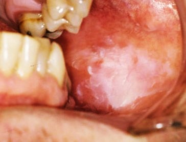
Leukoplakia
Leukoplakia can be defined as a white patch or plaque that cannot be characterized clinically or pathologically as any other disease. It is the most common oral premalignant lesion. Although leukoplakia can occur at any age, it often occurs in individuals under the age of 40. Its prevalence is six times more in smokers as compared to non-smokers. Leukoplakia results from chronic irritation of the mucous membranes by carcinogens; this irritation stimulates the proliferation of epithelial and connective tissue. Leukoplakia can be classified, based on clinical appearance into,
- Early/thin,
- Thick/homogenous,
- Granular/nodular,
- Proliferative verrucous, and
- Speckled leukoplakia types.

Thin early or thin leukoplakia appears as minimally elevated gray-white plaque with either well-defined or poorly defined borders. It is the initial stage of leukoplakia that gradually progresses to a thick, homogenous lesion with a leathery white fissured surface. However, all the initial lesions do not progress towards thick, homogenous lesion; instead, they progress to form the granular/nodular type with pebbly surface irregularities. A few lesions progress to a widespread multifocal lesion with a papillary surface. This uncommon variant of leukoplakia is called papillary verrucous ………………Content available in book……………….Content available in book……………….Content available in book……………….Content available in book……………….Content available in book……………….Content available in book……………….Content available in book…. The conditions that should be considered in the differential diagnosis of leukoplakia include the following,
- Aspirin burn,
- Chemical injury,
- Oral pseudomembranous and hyperplastic candidiasis,
- Frictional lesions,
- Oral hairy leukoplakia, leukoedema,
- Linea alba,
- Lupus erythematosus,
- Morsicatio buccarum,
- Papilloma and allied lesions,
- Mucous patches in secondary syphilis,
- Tobacco-induced lesions,
- Smoker’s palate (nicotinic stomatitis),
- White sponge nevus,
- Oral lichen planus (OLP), and
- Lichenoid reaction.
There are non-surgical as well as surgical treatment modalities for leukoplakia. Non-surgical treatment modalities might be considered in selected patients. Carotenoids (β-carotene, lycopene), vitamins [L-ascorbic acid (vitamin C), α-tocopherol (vitamin E), retinoic acid (vitamin A), and fenretinide], and bleomycin may be used in patients with oral leukoplakia. Surgical treatments are done more commonly. The most commonly preferred treatment options are surgical excision or CO2 laser therapy. In widespread lesions, photo-dynamic therapy may be considered. Another preferred treatment includes cryotherapy. In case of moderate to severe dysplasia, surgical excision is the treatment of choice.
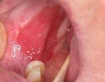
Erythroplakia
The term oral erythroplakia is used to describe a red plaque or macular lesion in the mouth for which a specific clinical diagnosis cannot be established. Lesions are named erythro-leukoplakia, leukoerythroplakia or speckled leukoplakia when red and white areas are associated or white patches are present over the red plaque. Most commonly affected areas were reported as the ……………Content available in book……………….Content available in book……………….Content available in book……………….Content available in book……………….Content available in book……………….Content available in book……………….Content available in book……………….Content available in book……………….Content available in book…. etiological factors in the development of erythroplakia are tobacco and alcohol use. Studies have reported a high prevalence of p53 mutations in premalignant oral erythroplakia. The differential diagnosis of erythroplakia includes,
- Oral atrophic candidiasis,
- Oral histoplasmosis,
- Oral tuberculosis,
- Atrophic OLP,
- Lupus erythematosus,
- Pemphigus,
- Pemphigoids,
- Amelanotic melanoma,
- Haemangioma,
- Telangiectasia,
- Lingual varies,
- Kaposi’s sarcoma,
- Early squamous cell carcinoma,
- Local irritation,
- Mucositis,
- Drug reaction,
- Median rhomboid glossitis, and
- Oral purpura.

The treatment of erythroplakia is primarily surgical excision owing to its high malignant transformation rate. Other treatment modalities include topical retinoic acid with systemic β-carotens; photodynamic therapy with methyl amino-levulinate; and cryosurgery or vaporization with carbon dioxide laser radiation. These approaches also include cessation of risk factors. Cases that have progressed to carcinoma, surgery (followed or not by radiotherapy), radio-therapy, and chemotherapy are recommended.
Palatal lesions of reverse smokers
Reverse smoking is highly damaging to the oral mucosa, specifically palatal mucosa which is directly exposed to the heat and smoke. It results in the formation of red areas, ulceration, pigmentation, excrescence patches, and palatal keratosis. The habit of reverse smoking is more in females than males in areas where this habit is prevalent. This may be because females want to keep their smoking habit a secret from their husbands and parents. Lesions associated with this habit range from palatal keratosis, excrescences, leukoplakia, ulcerations to frank malignancy. Term “palatal keratosis associated with reverse smoking” is a widely accepted finding associated with reverse smoking. The primary step in the treatment is the cessation of the habit. Surgical excision is indicated if there are indications of its progression to carcinoma.
Submucous fibrosis
Oral submucous fibrosis is a premalignant condition associated with the chewing of areca nut, an ingredient of betel quid, and is prevalent in South Asian populations. This condition can lead to squamous cell carcinoma, the risk which is further increased by concomitant tobacco consumption. The initial clinical feature of submucous fibrosis is burning sensation (usually occurs while chewing spicy food) and inflammation, which is followed by hypovascularity and fibrosis ……………Content available in book……………….Content available in book……………….Content available in book……………….Content available in book……………….Content available in book……………….Content available in book……………….Content available in book……………….Content available in book……………….Content available in book……………….Content available in book……………….Content available in book……………….Content available in book……………….Content available in book……………….Content available in book……………….Content available in book……………….Content available in book……………….Content available in book……………….Content available in book…. hyalinization extending into the submucosal tissues. Sometimes, dysplastic changes are observed in the epithelium.
As the condition advances with time, fibrous band forms in the buccal mucosa which restricts the mouth opening. The patient complaints of difficulty in mastication, speech, swallowing and maintaining oral hygiene. At this stage, the buccal mucosa becomes rubbery and difficult to retract or evert. With further advancement, the involvement of the floor of the mouth and tongue takes place that interferes with tongue movement. The involvement of palatal mucosa results in extensively blanched mucosa. The fibrosis may progress posteriorly towards the pharynx and may involve soft palate and uvula. In extreme cases, the patient faces difficulty in swallowing and may have loss of hearing due to blockage of the eustachian tube.
Cessation of the habit of betel nut chewing is the first step in the treatment of this premalignant condition. The patient should be educated and motivated to leave the habit as it may result in life-threatening consequences. Conventional therapies in the treatment of OSF are empirical and symptomatic in nature. The major targets of treatment can be summarized as anti-inflammatory, oxygen radical-scavenging and antifibrotic. Currently, intralesional steroids are the main treatment modality. These are injected into the fibrotic bands weekly for 6-8 weeks with regular monitoring of mouth opening. The oxygen radical-scavenging agents include lycopene, micronutrients and minerals are also used in the treatment. Fibrinolytic enzymes like hyalase, chymotrypsin and collagenase have shown beneficial results in lysis of collagen fibers. Other agents used include interferon gamma, turmeric, pentoxifylline, and CO2 laser.
Lichen planus
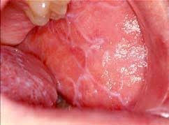
Lichen planus (LP) is a chronic autoimmune, mucocutaneous disease. It is believed to result from an abnormal T-cell-mediated immune response in which basal epithelial cells are recognized as foreign because of changes in the antigenicity of their cell surface. Oral lichen planus has been found to be associated with diseases and agents, such as viral and bacterial infections, autoimmune diseases, medications, vaccinations, and dental restorative materials. Lichen planus lesions are described using the six P’s; ……………Content available in book……………….Content available in book……………….Content available in book……………….Content available in book……………….Content available in book……………….Content available in book……………….Content available in book……………….Content available in book……………….Content available in book……………….Content available in book……………….Content available in book……………….Content available in book……………….Content available in book….

The typical oral manifestation of the LP is the presence of symmetrical white reticular lesions in the buccal mucosa, although diverse clinical features may be seen. Some patients report to the doctor with the sensitivity of the oral mucosa to hot or spicy foods, painful oral mucosa, red or white patches on the oral mucosa, or oral ulcerations. Patients with reticular lesions are often asymptomatic, but atrophic (erythematous) or erosive (ulcerative) oral LP is often associated with a burning sensation and pain. The definitive diagnosis of OLP depends on the histopathologic examination of the affected tissue. The classical picture of LP includes liquefaction of the basal cell layer accompanied by apoptosis of the keratinocytes and a dense band-like lymphocytic infiltrate at the interface between the epithelium and the connective tissue. Along with this, areas of atrophic epithelium where the rete pegs may be shortened and pointed (a characteristic known as sawtooth rete pegs) are seen. Focal areas of hyper-keratinized epitheli-um which give rise to the clinically apparent Wickham’s striae can be observed. Eosinophilic colloid bodies, also known as Civatte bodies, which represent degenerating keratinocytes, are often visible in the lower half of the surface epithelium. It should be remembered here that a similar histopathological picture may be seen in the lichenoid reaction to dental amalgam and drug. The differential diagnosis of oral LP includes,
- Chronic candidiasis,
- Morsicatio,
- White sponge nevus,
- Pemphigoid,
- Pemphigus,
- Discoid lupus erythematosus,
- Systemic lupus erythematosus,
- Hairy leukoplakia,
- Graft-versus-host disease,
- Chronic ulcerative stomatitis,
- Multifocal leukoplakia,
- Proliferative verrucous leukoplakia, and
- Squamous cell carcinoma.
There is no curative treatment for LP and the primary treatment is symptomatic. Corticosteroids are the drugs of choice in the treatment of lichen planus, due to their ability to modulate the inflammatory and immunological responses. Topical use and local injection of steroids have been used successfully for controlling lichen planus. Other drugs used in the treatment of lichen planus with good results include immunosuppressants such as cyclosporin and tacrolimus.
Epidermolysis bullosa
This is a dermatological disorder characterized by epithelial fragility that may manifest as blistering and erosions of the oral mucosa. There are three types of epidermolysis bullosa: simplex, junctional and dystrophic. Each type consists of several forms of disorders. The oral manifestations of the disease include marked frequency of oral and perioral blistering that leads to ulceration, scaring, and obliteration of the oral vestibule and microstomia. The oral lesions are primarily of junctional and dystrophic forms of the disease, which are characterized by bulla and vesicle formation following mild physical trauma. Junctional and dystrophic types are also associated with malignant transformation. The lingual mucosa of the oral cavity is ……………Content available in book……………….Content available in book……………….Content available in book……………….Content available in book……………….Content available in book……………….Content available in book……………….Content available in book……………….Content available in book……………….Content available in book……………….Content available in book…..
Actinic cheilitis of the lower lip
Actinic cheilitis is a chronic inflammatory lesion that primarily involves the lower lip. It results from excessive exposure to solar ultraviolet radiation; hence, the lower lip is commonly involved. The primary clinical features of actinic cheilitis are diffuse, poorly-defined atrophic, erosive, ulcera-tive or keratotic plaques. Other associated symptoms include dryness, scaly patches, swelling, transverse fissures, crusting, and blotching. The histopathological picture is characterized by varying degrees of keratosis, epithelial hyperplasia or atrophy, solar elastosis, and the presence or absence of dysplasia. Actinic cheilitis may transform into squamous cell carcinoma. The treatment options include cryotherapy, electrosurgery, topical retinoids, 5-fluorouracil cream, photo-dynamic therapy, carbon dioxide laser ablation, and vermilio-nectomy.
Discoid lupus erythematosus (DLE)
Lupus erythematosus is a chronic autoimmune disease. It has three forms: systemic, drug-induced, and discoid. The discoid variant affects the skin and may involve the mucosal surface of the lips and the oral cavity. Oral lesions may also manifest in approximately 20% of patients with systemic lupus. Malig-nant transformation of DLE into squamous cell cercinoma is usually observed on the lower lip, and in Caucasians. The histological features include hyperkeratosis, degeneration of the basal layer, and subepithelial lymphocytic infiltration. Topical or systemic corticosteroids are usually recommended for management of discoid lupus erythematosus.
Conclusion
Oral premalignant lesions (OPLs) are changes in the tissues of the mouth that have the potential to progress to oral cancer if left untreated. These lesions are typically detected during routine dental examinations or due to symptoms such as persistent sores, pain, or changes in the color or texture of the oral mucosa. So, regular dental check-ups and awareness of changes in the oral cavity play a significant role in early identification and intervention. Many conditions can be treated by use of topical or systemic medications to treat underlying conditions or reduce dysplasia. Sometimes excision of the lesion is necessary to prevent progression to malignancy. Addressing risk factors such as tobacco and alcohol use, improving oral hygiene, and protecting against UV exposure are done to prevent malignancy.
References
References are available in the hardcopy of the website “Textbook of basic sciences for MDS students”.
Periobasics: A Textbook of Periodontics and Implantology
The book is usually delivered within one week anywhere in India and within three weeks anywhere throughout the world.
India Users:
International Users:

