Introduction to vascular supply and innervation of the gingiva
The orofacial structures have a very rich vascular supply. The periodontium is a highly vascular and innervated structure in the orofacial region. There is a network of blood vessels that supply and drain teeth and their supporting tissues. A very important artery that supplies the maxillary and mandibular teeth and their associated periodontium is the internal maxillary artery. In-depth knowledge of this artery is important before we go ahead with the arterial supply of mandibular and maxillary gingiva.
Internal maxillary artery
The maxillary artery or internal maxillary artery is one of the two terminal divisions of the external carotid artery. The second terminal branch is the superficial temporal artery. The maxillary artery is the largest branch of the external carotid, arising under the cover of the parotid gland, just above the posterior auricular artery. It is hidden behind the zygomatic arch. After its origin, it runs between the two heads of the lateral pterygoid muscle to penetrate the pterygopalatine fossa. It supplies the deep structures of the face.
The maxillary artery is divided into three portions by its relation to the lateral pterygoid muscle:
- First (mandibular) part: posterior to lateral pterygoid muscle (five branches).
- Deep auricular artery (enters squamotympanic fissure).
- Anterior tympanic artery (enters squamotympanic fissure).
- Middle meningeal artery (enters foramen spinosum).
- Accessory meningeal artery (enters foramen ovale).
- Inferior alveolar artery (enters the mandibular foramen).
- Artery to mylohyoid.
- Second (pterygoid or muscular) part: within lateral pterygoid muscle (five branches).
- Anterior deep temporal branches.
- Posterior deep temporal branches.
- Pterygoid branches.
- Masseteric artery.
- Buccinator artery.
- Third (pterygopalatine) part: anterior to lateral pterygoid muscle (six branches including terminal branch).
- Posterior superior alveolar artery.
- Infraorbital artery (enters the inferior orbital fissure).
- Artery of the pterygoid canal.
- Pharyngeal artery (enters palatovaginal canal).
- Greater (descending) palatine artery (enters the greater palatine foramen).
- Sphenopalatine artery – terminal branch (enters the sphenopalatine foramen).
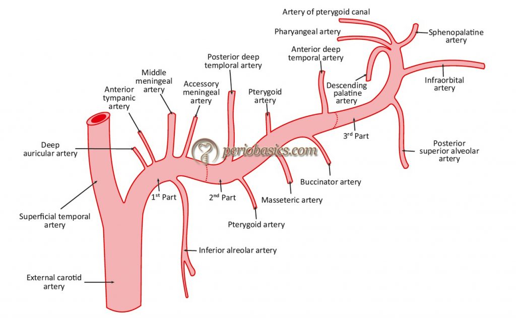
Branches from the first part
Deep auricular artery:
It is the first branch of the maxillary artery that originates from its first part. It passes through the bony or cartilaginous wall of the external acoustic meatus to supply the skin of that canal and part of the tympanic membrane. Sometimes, it also contributes a vessel to the temporomandibular joint.
Anterior tympanic artery:
It is the second branch of the first part of the maxillary artery. The vessel derived from this artery passes through the petrotympanic fissure to supply the lining of the middle ear and accompanies the chorda tympani in its course.
Middle meningeal artery:
The middle meningeal artery enters the skull through the foramen spinosum. It gives off branches for the dura mater surrounding the trigeminal ganglion. This is the main source of blood for the dura mater
Accessory meningeal artery:
This artery passes upwards through the foramen ovale to supply the trigeminal ganglion and the dura mater of Meckel’s cave and the middle cranial fossa. It also usually supplies the medial and superior head of the lateral pterygoids.
Inferior alveolar artery:
This branch of the maxillary artery runs downwards and forwards towards the inferior alveolar nerve, to meet the nerve at the mandibular foramen. The artery runs further anteriorly in the mandible, supplying the pulps of the mandibular teeth (with its dental branches) and the body of the mandible. Its other branch, the mental branch, emerges from the mental foramen and supplies the nearby lip and skin.
Branches from the second part
Anterior and posterior deep temporal branches:
The deep temporal arteries (anterior and posterior) are branches from the second part of the maxillary artery. They ascend between the temporalis muscle and the pericranium supplying the overlying muscle.
Pterygoid branches:
This artery supplies blood to pterygoid muscles.
Masseteric artery:
This narrow artery carries the blood flow to the masseter muscle.
Buccinator artery:
It runs obliquely forward, between the medial pterygoideus and the insertion of the temporalis muscle, to the outer surface of the buccinator muscles, to which it supplies.
Branches from the third part
Posterior superior alveolar artery:
The posterior superior alveolar artery is a branch of the third (peterygopalatine) part of the maxillary artery. Descending upon the tuberosity of the maxilla, it divides into numerous branches, some of which enter the alveolar canals, to supply the molar and premolar teeth and the lining of the maxillary sinus, while others are continued forward on the alveolar process to supply the gingiva.
Infraorbital artery:
It passes forwards through the inferior orbital fissure, along the floor of the orbit and the infraorbital canal to emerge with the infraorbital nerve on the face through infraorbital foramen.
Artery of the pterygoid canal:
The pterygoid canal, also known as the Vidian canal. It is a foramen in the base of the skull, which is located in the sphenoid bone, inferomedial to the foramen rotundum. It transmits the Vidian artery and Vidian nerve from the middle cranial fossa to the pterygopalatine fossa. It supplies the upper part of the pharynx, and sends a small division into the tympanic cavity to anastomose with the tympanic arteries.
Pharyngeal artery:
It runs backwards through the canal related to the inferior aspect of the body of the sphenoid bone (palatovaginl canal). It supplies structures such as the pharynx, roof of the nose, the auditory tubes and the tympanic cavity.
Greater (descending) palatine artery:
It runs downwards in the greater palatine canal to emerge on the posterolateral part of the hard palate through the greater palatine foramen. It then runs forwards near the lateral margin of the palate to reach the incisive canal through which some of its branches enter the nasal cavity. The branches of this artery supply the palate and the gums. In the greater palatine canal, it gives off the lesser palatine branch that emerges on the palate through the lesser palatine foramen and runs backwards into the soft palate and tonsil.
Sphenopalatine artery:
The sphenopalatine artery (formerly known as the nasopalatine artery) passes through the sphenopalatine foramen into the cavity of the nose, at the back part of the superior meatus. Here it gives off its posterior lateral nasal branches which spread forward over the conchae and meatuses, anastomosing with the ethmoidal arteries and the nasal branches of the descending palatine, and assist in supplying the frontal, maxillary, ethmoidal, and sphenoidal sinuses. This artery is also referred to as the artery of epistaxis due its common involvement in cases of nose bleeds. It is a major contributor to the rich arterial plexus known as Kiesselbach’s plexus on the anteroinferior nasal septum.
Arterial supply of mandibular and maxillary gingiva
The mandibular teeth and their supporting structures are supplied by branches of inferior alveolar (dental) artery, including mental, sublingual and buccal arteries. The inferior alveolar artery is a branch of the internal maxillary artery. Near its origin, the inferior alveolar artery has a lingual branch which supplies the tongue and adjacent mucous membrane. The inferior alveolar artery descends posterior to ……………………..Contents available in the hard-copy of the website………… Contents available in the hard-copy of the website……..

Periobasics A Textbook of Periodontics and Implantology
The book is usually delivered within one week anywhere in India and within three weeks anywhere throughout the world.
India Users:
International Users:
The arterial supply of the maxillary teeth and their supporting structures is by the posterior superior alveolar artery, infraorbital artery, the greater palatine artery and the sphenopalatine arteries. The posterior superior alveolar artery gives branches over the maxillary tuberosity supplying the maxillary teeth in the molar region, alveolar bone and the mucosa. These branches anastomose with the branches of the facial artery. The anterior superior alveolar artery, which is a branch of the infraorbital artery, curves through the canalis sinuosus following the rim of the anterior nasal aperture supplies the anterior teeth in the canine and incisor region. On the palatal aspect, the greater palatine artery supplies the palatal gingiva. In the anterior region the nasopalatine artery supplies the palatal gingiva.
The gingiva receives its arterial supply mainly from three sources: the supraperiosteal arterioles, the vessels from the periodontal ligament and arterioles emerging from the crest of the interdental septa. Out of these three sources, the gingiva is primarily supplied by the periosteal arterioles that course over the alveolar process. These arterioles supply a capillary plexus that course below the gingival epithelia. They form anastomoses with capillaries from the periodontal ligament and alveolar bone 191, 192. Fine capillary loops can be seen extending into the papillary layer of the epithelium from the capillary plexus 193. The approximate diameter of the blood vessels in this plexus is 40 μm, indicating that these are mainly venules. The capillary plexus beneath the junctional epithelium lacks distinct capillary loops 97, 194, 195. However, during inflammation, these capillaries enlarge and acquire varicosities 196, 197. These capillaries subjacent to the junctional epithelium are a major source of the gingival crevicular fluid and the inflammatory cells that migrate into the lamina propria and then into the junctional epithelium during inflammation 198, 199.
Venous and lymphatic drainage
The venous and the lymphatic drainage of gingiva is closely associated with the arterial supply. In the maxilla, the gingival lymphatic vessels drain into the deep cervical lymph nodes. In the mandible, they drain into the mental, submandibular and the cervical lymph nodes. These lymphatic vessels return the fluid and filterable plasma components to the blood via thoracic duct.
Innervation of the gingiva
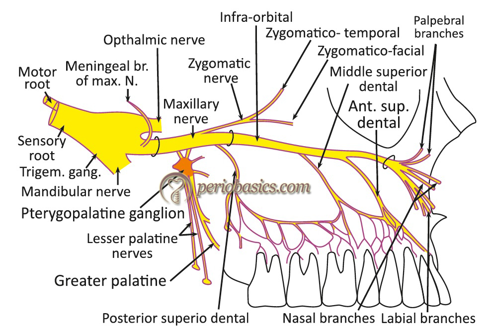
The gingiva in various regions of the oral cavity is supplied by the second and the third division of the trigeminal nerve. The nerve supply of gingiva follows the course of vascular supply. In the maxilla, the gingiva is supplied by the posterior, middle and the anterior superior alveolar nerve, branches of the infraorbital nerve, the greater palatine nerve, and nasopalatine nerve. The middle superior alveolar nerve is present in around 80% of individuals. The buccal nerve supplies variably in the buccal molar region.
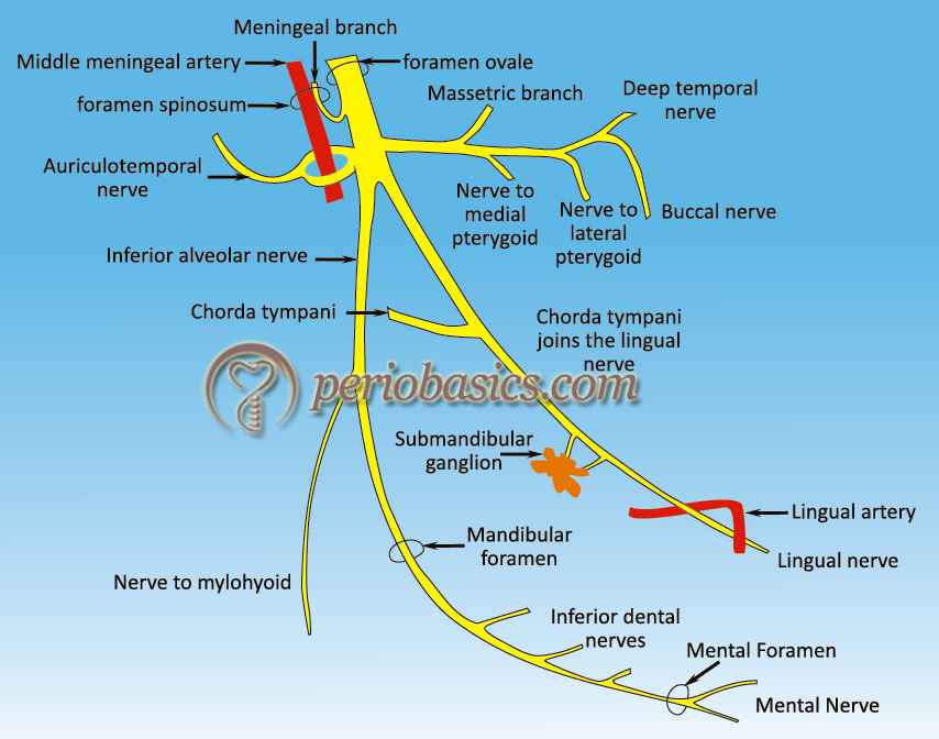
In the mandible, the gingiva is largely supplied by the inferior alveolar nerve. The buccal nerve supplies buccal gingiva in relation to the molars and premolars. The branches of the lingual nerve supply the gingiva on the lingual aspect of all lower teeth. Most of the nerve endings in gingiva terminate within the lamina propria. Only a few nerve endings are present in the epithelium. Various neural terminal endings present in gingiva include Meissner corpuscles, Krause type end bulbs, and encapsulated spindles.
Conclusion
Thorough knowledge of the vasculature and innervation of the periodontium is necessary from the clinical point of view. Various surgical procedures are carried out in his area for various purposes. A thorough understanding of the anatomical structures in this area is essential before making any surgical intervention in this area.
References
References are available in the hardcopy of the website.
Periobasics A Textbook of Periodontics and Implantology
The book is usually delivered within one week anywhere in India and within three weeks anywhere throughout the world.