Introduction to guided bone regeneration
Periodontal regeneration has been a field of research for quite a long time now. Although, complete periodontal regeneration is still to be achieved; we have succeeded in developing techniques which enhance regeneration. These techniques include root surface biomodification, guided tissue regeneration, application of fibrin-rich, and platelet-rich plasma in periodontal regeneration and guided bone regeneration. In the present discussion, we shall study in detail the principles of guided bone regeneration (GBR).
This procedure is used to increase bone volume in the areas with bone resorption due to long-standing loss of tooth/teeth. The placement of fixed partial denture in these areas results in an un-esthetic appearance due to reduced bone and tissue volume (ridge defect). Another important indication of GBR is to increase the bone volume in areas with deficient bone for dental implant placement. The prime requirement for implant placement is the availability of bone. In long-standing edentulous areas, usually there is bone resorption, which makes it difficult to place implants and also affects their long-term stability. GBR procedure increases the bone volume, thus facilitating implant placement as well as its long-term stability.
Guided bone regeneration is based on the concept of ………. Contents available in the book ………. Contents available in the book ………. Contents available in the book ………. Contents available in the book ………..

Periobasics: A Textbook of Periodontics and Implantology
The book is usually delivered within one week anywhere in India and within three weeks anywhere throughout the world.
India Users:
International Users:
Pioneer work on guided bone regeneration
One of the earliest research papers on GBR using the application of barriers in bone healing was published by Murray et al. (1957) 1, where they treated femoral bone defects in dogs that were protected by rigid plastic cages. After an appropriate healing period, it was noted that the defect got filled with bone consistent with the original cortex. They suggested that the soft tissue which grows at a faster rate than bone hinders bone formation in the healing area and should be prevented to promote bone formation. Further, they stated that only three conditions were necessary for the new growth of bone: the presence of a blood clot, preserved osteoblasts, and contact with living tissue. In 1980’s research work done by Karring and Nyman highlighted the concept of GTR which acted as a foundation for GBR.
In 1980’s, Dahlin et al. (1988, 1989, 1990) 2-4 tried to reconstruct large atrophic bone defects in maxilla and mandible. Their focus was to re-establish bone volume in areas where due to long-standing edentulism, a lot of bone resorption had already taken place. Urist et al. (1963) 5 had already shown that for bone formation, the rate of osteogenesis extending inward from the adjacent bone ends must exceed the rate of fibrogenesis growing in from the surrounding muscle or connective tissue. Dahlin et al. (1988) 2 in an animal experiment evaluated the outcome of GBR procedure. They created bilateral through and through defects in ramus of 30 Sprague-Dawley rats. On one side, the defect was covered with a porous polytetrafluoroethylene (PTFE) membrane (Gore-Tex®) and the other side served as a control, without a membrane covering. The macroscopic and histological examination of the specimens after ………. Contents available in the book ………. Contents available in the book ………. Contents available in the book ………. Contents available in the book ………..
The application of GBR procedure around implants was investigated in another study by Dahlin et al. (1989) 3, where they placed 30 ”commercially pure” 10-mm titanium implants in the tibia of 15 adult rabbits. Three to four threads of each implant were left exposed. A porous polytetrafluoroethylene (PTFE) membrane (Gore-Tex®) was placed over the test fixtures, covering the threads and 5 to 8 mm of the adjacent bone. The control fixtures were not covered by a membrane. The en bloc specimens were taken after healing periods of 6, 9, and 15 weeks and evaluated grossly and histologically. The results of the study showed that all exposed threads of the titanium implants were covered with newly formed bone at a uniform thickness, even as early as 6 weeks. The new bone formation was also seen in the control areas, but was much lesser than that the test areas. This study further highlighted that by placing a barrier membrane, ingrowth of osteoprogenitor cells in the area of healing is enhanced causing new bone formation. Later on, various animal studies investigated the efficacy of application of resorbable and non-resorbable membranes in maxillary and mandibular bone defects in GBR 6-9.
Ridge augmentation for implant placement by using resorbable collagen membrane was investigated in one study 10. In this study, collagen membranes were used to augment alveolar ridge deficiencies. Following treatment, 75% of the defects treated with collagen membranes demonstrated sufficient width for implant placement. In another study, collagen ………. Contents available in the book ………. Contents available in the book ………. Contents available in the book ………. Contents available in the book ………..
Indications of guided bone regeneration
In the majority of cases, GBR procedure is used to increase the bone volume in bone deficient ridges. Following are the potential indications of GBR 12,
- Local alveolar ridge deficiencies (horizontal or limited vertical).
- Osseous fill around immediate implants.
- Dehiscence and fenestrations associated with implants.
- Bone defects associated with failing implants.
- Residual bone lesions.
- To aid in repair of sinus membrane perforations.
Principles of guided bone regeneration
GBR and GTR share the same principle of placing a barrier membrane for selective growth and proliferation of desired cell population which causes regeneration of lost periodontal structures. Following are the basic requirements for GBR,
Cell exclusion:
The cell population that hinders the regeneration process such as gingival fibroblasts and/ or epithelial cells should be prevented form reaching the area of healing. This causes the formation of fibrous connective tissue which is not desirable. Barrier membrane is placed to prevent ingress of undesired cells into the wound area. There are various barrier membranes available, both resorbable and non-resorbable. A detailed description of barrier membranes has been given in chapter 66 “Guided tissue regeneration”.
Tenting:
The area where bone formation is desired must be secured by placing the membrane. The membrane is carefully placed in such a way that space is created beneath the membrane which completely ………. Contents available in the book ………. Contents available in the book ………. Contents available in the book ………. Contents available in the book ………..
Scaffolding:
The tented space provided by the membrane is occupied by fibrin clot that acts as a scaffold. It acts as a framework for cell migration and proliferation. The chemical mediators released by platelets in the clot promote cell migration in the wound area. These include inflammatory cells such as monocytes and PMN’s and progenitor cells. In the case of GBR, progenitor cells migrate from adjacent bone or bone marrow.
Stabilization:
It is the prime requirement for regeneration. The clot beneath the membrane should be protected from any kind of disruption due to movement of the membrane. The membrane is therefore often, but not always, stabilized by sutures/ mini bone screws/ bone tacks. In situations where sutures/ mini bone screws/ bone tacks cannot be placed, the membrane is simply tucked beneath the margins of the flap, so that after suturing the membrane is stable in its position.
Framework:
In areas such as dehiscence or fenestration, the membrane collapses and cannot maintain the space. In such areas, the membrane should be supported by a framework to prevent it from collapsing. Bone grafts such as autografts, allografts or xenografts may be placed in such areas which act as a framework, preventing membrane collapse. Stiffer membranes such as titanium-reinforced membranes can also be used in such areas.
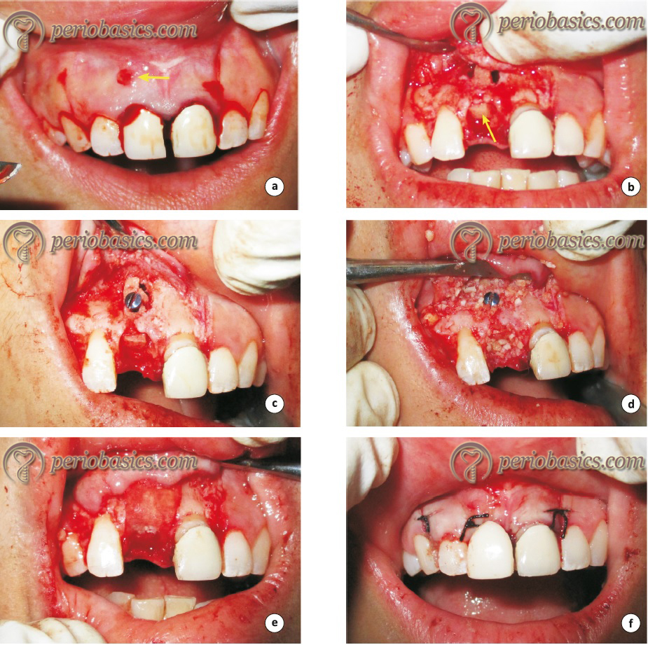
Clinical procedure
Although, the surgical technique may vary according to the clinical presentation of the defect, but following are the basic surgical steps followed during GBR,
- After achieving profound local anesthesia, an incision is made in such a way that it is not placed directly over the defect. This is done so that the closure line should not be directly over the defect but away from the defect site. By doing this, we can achieve effective closure of the mem-brane. Vertical releasing incisions are given wherever indicated.
- A full-thickness flap is then reflected 2-3 mm beyond the margins of the defect. After this, an incision is made through the periosteum to make it a partial-thickness flap. The partial-thickness flap is then raised using blunt dissection. The purpose of continuing with partial thick-ness flap is to achieve primary closure without tension because periosteum is not stretchable.
- The defect is then debrided of all the granulation tissue and is carefully examined.
- The flap is trimmed and epithelium and tissue ………. Contents available in the book ………. Contents available in the book ………. Contents available in the book ………. Contents available in the book ………..
- After this, the barrier membrane is taken out of its packing and is trimmed in such a way that its margins extend 2-3 mm beyond bony margins of the defect. When using collagen membrane, it may be hydrated in normal saline before using to improve its malleability.
- Small cortical perforations are then made with the help of ½ round bur in the bone to induce bleeding points. These act as sites of egress of osseous progenitor cells into the defect.
- The bone graft is then placed in the defect to support the tented membrane. Tenting screws alone or in conjunction with bone grafts can also be used for this purpose. If corticocancellous graft is used, it is stabilized in position with the help of screws.
- The membrane is then placed and adapted to the defect site. If the membrane is stable in its place, no attempt is made to fix it, but if it is not stable, then pins, bone screws, or tacks may be needed to assist membrane stability.
- The surgical site is then closed with the help of silk, vicryl or e-PTFE sutures.
Romanos (2010) 13 in an article discussed following guidelines while raising a mucoperiosteal flap for GBR procedure during ridge augmentation,
- The flap raised for GBR procedure has to be a mucoperiosteal flap and it must be raised sufficiently to achieve complete coverage of the augmented site. It should be raised over the mucogingival junction in an apical direction upto at least 10 mm.
- To evaluate the tension in the flap during coverage of the augmentation site, hold the flap (but not pull it) in a coronal direction with conventional dental forceps (surgical forceps may lead to flap perforations). If the flap is under tension while coverage of augmented site, an incision is made through the periosteum. The incision is made with ………. Contents available in the book ………. Contents available in the book ………. Contents available in the book ………. Contents available in the book ………..
- The blade should cut the tissue in a level apical to the mucogingival junction. The incision is not made in the keratinized mucosa to avoid any flap perforation.
- The flap is again pulled and checked for tension-free flap advancement. In case of insufficient closure, a deep incision is made in the muscle layer and vertical incisions may be extended more apically. Muscle release can be performed using dissection scissors.
- To confirm appropriate tension-free closure, make sure that the buccal flap margin covers the lingual or palatal site at least upto 3-5 mm. If this overlapping does not occur, there is tension in the flap, which means the closure is insufficient.
Biological aspect of GBR
Healing after GBR procedure is a complex process where sequential cellular and molecular events take place to generate new bone. There is a précised series of events that take place during post-operative healing. These events may be modulated by systemic and local factors, and disruption of these orderly events may cause healing problems. Formation of the blood clot is accompanied by an inflammatory response, where proinflammatory cytokines such as interleukin-1 (IL-1), IL-6, and particularly tumor necrosis factor-α, initiates the bone healing cascade.
Various growth factors, particularly the transforming growth factor-β (TGF-β) superfamily, including bone morphogenetic protein (BMPs), as well as platelet-derived growth factors (PDGF), fibroblast growth factors (FGFs) and insulin-like growth factors (IGFs), orchestrate crucial events for osteogenesis. Another key factor during healing after GBR procedure is angiogenesis. Vascular endothelial growth factor (VEGF) plays a key role in angiogenesis along with ………. Contents available in the book ………. Contents available in the book ………. Contents available in the book ………. Contents available in the book ………..
A negative feedback control is also required to regulate the functioning of growth factors and other molecules. Growth factor inhibitors and various BMP antagonists are released into the extracellular compartment including noggin, sclerostatin, follistatin, etc. Other inhibitory mechanisms include receptor inhibition of some members of the TGF-β superfamily that has been related to a pseudo-receptor defined as BAMBI (BMP and activin membrane-bound inhibitor) 14, and intracellular inhibition by the activation of I-Smads 15, 16.
Another important factor during wound healing is scaffold used for new bone formation. The primary requirements of a scaffold include,
- It must provide the correct anatomic geometry to define and maintain the space for tissue regeneration.
- The scaffold must provide temporary mechanical load-bearing within the tissue defect.
- The scaffold should enhance the regeneration and should be able to carry biologically active molecules in the healing area.
- Various bone substitutes and barrier membranes have been explained in detail in “Bone grafts in periodontics” and “Guided tissue regeneration”, respectively.
GBR in implantology
GBR is a quite promising technique to regain the bone volume. In implantology, GBR is applied in following ways: One-stage lateral augmentation in which the procedure is done for treatment of dehiscence or fenestration defects during implant installation and Two-stage bone augmentation procedure in which bone augmentation is done prior to implant placement.
One-stage lateral augmentation:
During implant placement, dehiscence or fenestration defect is a common surgical complication. This is because of insufficient bone volume at the surgical site. Once the defect is identified, GBR procedure is done to cover the exposed implant surface with new bone. The procedure involves placement of bone graft over the involved area and putting a barrier membrane to cover the defect with 2-3 mm extensions on all the sides. The membrane should be stable in its position so that the healing remains uninterrupted.
Two-stage bone augmentation procedure:
It is done in areas where during implant placement, primary stability cannot be achieved. So, increasing the bone volume before implant placement becomes essential. Two different situations are encountered here: long-standing edentulous span with deficient ridge and tooth extraction socket that shall leave behind the insufficient bone. In the former situation bone augmentation is done following the surgical technique already explained in the previous sections. In the latter case, the involved tooth is extracted, the socket is debrided and cleaned and bone graft is placed in the extraction socket. After packing the bone graft in the socket, it is covered by a barrier membrane and sutures are placed to close the area. Research has demonstrated that ………. Contents available in the book ………. Contents available in the book ………. Contents available in the book ………. Contents available in the book ………..
In insufficient edentulous ridge, lateral augmentation has been shown to be successful and predictable, but results with vertical augmentation are controversial. This is because of the reason that the vertical augmentation procedure is technique sensitive and key factors for the success of this procedure are yet to be identified, making this procedure unpredictable 22.
GBR in immediate implant placement
The shape of the circumference of the tooth at bone level is different from that of the implant, especially in the case of molars. When an immediate implant is planned in an extraction socket, all the surfaces of the implant in the coronal portion may not have a direct contact with socket walls. After achieving primary stability, the distance of the implant surface from the socket walls is evaluated. The jumping distance (read in “Biological aspect of dental implants”) of the bone is approximately 2 mm. So, if the distance is more than this, we need to go for GBR procedure. The bone graft and barrier membrane are placed, and the area is closed with ………. Contents available in the book ………. Contents available in the book ………. Contents available in the book ………. Contents available in the book ………..
Factors affecting outcome of guided bone regeneration
There are many factors which directly or indirectly affect the outcome of a GBR procedure. GTR and GBR procedures share many factors in common, including patient factors, clot stabilization, defect morphology, membrane properties and fixation and innate regenerative potential of the patient. These factors have already been discussed in detail in “Guided tissue regeneration”. On the basis of clinical research, many authors have enumerated the factors affecting the outcome of GBR therapy. Hermann and Buser (1996) 24 discussed five surgical factors that are required to achieve predictable results with GBR procedures:
1. Use of an appropriate membrane;
2. Achievement of primary soft tissue healing;
3. Creation and maintenance of a membrane-protected space;
4. Close adaptation and stabilization of the membrane to the surrounding bone; and
5. Sufficiently long healing period.
Wang and Boyapati (2006) 25 have described “PASS” principles for predictable bone regeneration during GBR procedure. These include primary wound closure, angiogene-sis, space creation/maintenance, and stability of both the initial blood clot and implant fixture (PASS). Wound stability and angiogenesis play a key role in bone formation. Along with this, many investigators have advocated the necessity of primary closure following implant placement to ensure predictable GBR outcomes 26-28. Other than common factors shared by both GTR and GBR procedures, let us now discuss in detail, factors specifically affecting the success of GBR,
Ridge morphology:
Ridge morphology is an important factor affecting the outcome of GBR procedure. The significance of ridge morphology was evaluated by Park et al. (2009) 29, who in a retrospective study, analyzed conventional tomograms to estimate the prognostic value of the cross-sectional ridge morphology on the clinical outcome of the GBR procedure in buccal dehiscence defects in 23 single implant sites. They reported that ridge width did not have a significant effect on the regenerative outcome, but cross-sectional ………. Contents available in the book ………. Contents available in the book ………. Contents available in the book ………. Contents available in the book …………
Incision location:
The vascular supply of the ridge mucosa is an important factor to be considered. Because an avascular zone located over the edentulous ridge is about 1 to 2 mm wide, it may be inferred that mid-crestal incisions on the edentulous ridge with a possible vertical incision on the mesial aspect of the flap seem to yield the most anatomic potential for success 30. It has been shown in human studies that a crestal incision causes less edema, inflammation, and pain as compared to vestibular or mucobuccal fold incisions. Other benefits of crestal incision include reduced flap necrosis and reduced risk of membrane exposure 31.
Tension in the flap:
While doing ridge augmentation, the surface area of the ridge is increased. The soft tissue must completely cover this augmented ridge passively. The releasing incisions and incision in the periosteum make the flap flexible as discussed in the surgical technique. Any undue tension in the flap may cause membrane exposure and other post-operative complications, including suture line opening and infection 32. The methods suggested to alleviate tension over the suture line include overlapping the flap, positioning the flap laterally, tissue grafting, or using a combination of mattress sutures and interrupted sutures 32.
Kfir et al. (2007) 32 introduced the minimally invasive GBR technique in which they used sequential balloon inflations to create space for membrane placement. Initially, a vertical incision is made mesial to the augmentation zone. A miniature chisel is used to elevate the periosteum initially, followed by a series of sequential balloon inflations. At the end of the procedure, a tunnel with adequate space for membrane insertion, decortication, and ………. Contents available in the book ………. Contents available in the book ………. Contents available in the book ………. Contents available in the book ………..
Decortication (Intra-marrow penetration):
It is a procedure in which intentional holes are drilled in the cortical plate into the cancellous bone so that osteoprogenitor cells can reach the area of healing. Decortication may also improve the physical bond between grafted bone and a recipient site 33, 34. Although, scientifically the concept of decortication holds good, but clinical data on this procedure has conflicting results regarding its ability to enhance new bone formation 34, 35.
Review of literature on long-term results of GBR and other ridge augmentation procedures
The GBR procedure is primarily done for ridge augmentation to provide an adequate bone support to the implant. Various studies have investigated the long-term success rate of implants placed in the regenerated bone.
A study investigated failure and success rates of 626 implants either placed in the regenerated alveolar bone or treated with GBR to rebuild bone over implant fenestrations or dehiscences. The authors reported that cumulative success rate of implants in function in regenerated bone for 6-51 months was round 93.8% 36. In another study, a total of 38 implants placed in 7 patients were investigated for their long-term success. Out of these 38 implants, 21 implants were placed in conjunction with GBR using a polylactic, polyglycolic acid membrane and 17 were placed into the host bone without the need for regeneration therapy. After an average of 25 months following the incorporation of fixed reconstructions, a 100% survival rate was found for both test and control implants and no significant difference in marginal bone levels was recorded between the two groups 37.
A systematic review 38 was done to identify the most successful technique(s), which provided the necessary alveolar bone to place a dental implant and its long-term survival. Various bone augmentation techniques were investigated, including guided bone regeneration (GBR), onlay/veneer grafting (OVG), combinations of onlay, veneer, interpositional inlay grafting (COG), distraction osteogenesis (DO), ridge splitting (RS), free and vascularized autografts for discontinuity defects (DD), mandibular interpositional grafting (MI), and socket preservation (SP). A total of 2,620 implants placed in the augmented alveolar ridge were included from various studies, with follow-up ranging from 5 to 74 months. The implant survival rate was ………. Contents available in the book ………. Contents available in the book ………. Contents available in the book ………. Contents available in the book ………..
In another review, the success of different surgical techniques for the reconstruction of edentulous deficient alveolar ridges was evaluated. The study included 10 consecutively treated patients with a mean follow-up of at least 12 months after commencement of prosthetic load. The surgical procedures analyzed included onlay bone grafts, sinus floor elevation via a lateral approach, Le Fort I osteotomy with interpositional grafts, split ridge/ridge expansion techniques, and alveolar distraction osteogenesis. The authors concluded that it was difficult to demonstrate that one surgical procedure offered better outcomes than another because every procedure has its advantages and disadvantages. Priority should be given to those procedures which are simpler and less invasive, involve less risk of complications, and reach their goals within the shortest time frame 39.
Conclusion
The available evidence clearly shows the predictability of the GBR procedure in the regeneration of bone in deficient alveolar ridges both in height and width. Although, this procedure is technique sensitive, but if properly done, desirable results can be achieved. Long-term results of many studies have demonstrated that the survival rate of implants placed in augmented bone is high and comparable with implants placed in the native bone. Studies have indicated more complications while achieving vertical ridge augmentation rather than horizontal ridge augmentation. More clinical trials are required to test the validity of GBR in vertical and horizontal bone augmentation. Also, low level of evidence is available to substantiate the validity of GBR in bone regeneration in extreme bone resorption cases and with bone regeneration involving maxillofacial surgeries. In the future, we shall see more research in this field because GBR has become an inseparable part of implant placement in deficient alveolar bone.
References
References are available in the hard-copy of the website.
Periobasics: A Textbook of Periodontics and Implantology
The book is usually delivered within one week anywhere in India and within three weeks anywhere throughout the world.