Introduction to dental calculus
Although, dental plaque is considered as the primary etiological factor in the development of periodontal diseases; the presence of dental calculus is also of great concern to the clinicians because it facilitates plaque formation by providing the surface for its formation and keeps it in close contact with the gingival tissue. Furthermore, the plaque deposited on the surface of calculus in the subgingival areas is very difficult to clean by self-performed oral hygiene measures used by the patient. Hence, complete removal of the calculus and necrotic cementum from the root surfaces of the teeth by oral health care providers is essential to achieve periodontal health.
Definition
Calculus can be defined as a hard concretion that forms on the teeth or dental prosthesis through the calcification of bacterial plaque 1.
Historical aspect
The belief that calculus in some way or the other is related to periodontal diseases can be traced back to the 10th century. The discovery of toothpicks of gold and silver in Sumerian tombs dating as back as 3500 BC implies the importance given to oral hygiene and the removal of tooth deposits throughout the ages. Perhaps the first formal association between dental deposits and oral disease can be found in the writings of Hippocrates (460-377 BC), the Greek physician who founded modern medicine. He noted the deleterious effects of ‘pituita’ (calculus, which insinuated itself, on the roots of the teeth) on the teeth and gums.
Albucasis (936-1013 AD), an Arabian physician and surgeon, was the first to clearly enunciate the relationship between calculus and periodontal diseases. He emphasized on the need for removal of these deposits around the teeth. Furthermore, he designed a set of scaling instruments for the purpose of removing calculus in patients afflicted with periodontal diseases.
In 1535 Paracelsus, a German physician and alchemist introduced the term ‘tartar’ as a designation to a variety of stony concretions that form in humans, noting their physical comparability to the deposits that develop at the bottom of wine casks. He observed that these ‘tophi’ could be found around the teeth, in the urinary bladder, in the gallbladder, and in gouty joints. Paracelsus looked upon tartar as the principal cause of certain maladies, which he termed “tartaric diseases”. Pierre Fauchard in 1728 referred this slime layer as “a substance which accumulates on the surface of the teeth and which, when left there, becomes a stony crust of more or less considerable volume”. He further stated that due to the deposition of this foreign substance around the teeth, gum diseases are initiated because of which in due course of time teeth are lost.
With the introduction of microscopes and other bacterial identification techniques, our understating regarding the etiopathogenesis of periodontal diseases improved significantly during the nineteenth century. Dental plaque was established as the primary etiological factor for periodontal diseases. Dental calculus was just considered as a ‘fossilized remnant of minor significance’ in the etiopathogenesis of periodontal diseases. However, Mandel (1974) 2 asserted that ignoring calculus for its etiopathogenic significance was a premature relegation since the calcified deposits do contribute to the development of pathologic conditions.
Epidemiological studies done on calculus
Various epidemiological studies have been done to find out supra- and subgingival distribution of calculus and its correlation with the associated periodontal status. In one study, Ainamo (1970) 3 examined 154 army recruits (19-22 years of age) and investigated if any correlation existed between calculus (supra and subgingival) and gingivitis. The results of the study demonstrated a high correlation between the presence of calculus and gingivitis. In another study, Alexander (1971) 4 examined the distribution pattern of plaque, supra-, and subgingival calculus and gingival inflammation in 200 dental students and 200 dental clinic patients. The results of the study demonstrated that the highest prevalence of gingival inflammation was associated with interdental papillae and lowest with buccal margins. This pattern of gingival inflammation distribution coincided with the highest prevalence of subgingival calculus on the interproximal surface and the lowest on the buccal surfaces. However, in this study, it was observed that the distribution pattern for gingival index and plaque was much closer than the distribution pattern for gingival index and subgingival calculus. Thus, it was suggested that plaque had a much closer association with gingival inflammation than calculus. These findings were supported by another study done by Buckley (1980) 5 who examined 300 subjects aged between 15-17 years.
Studies have also been done to evaluate the effects of calculus removal on periodontal health. In a study, Tagge et al. (1975) 6 evaluated the effects of scaling, root planing and personal oral hygiene versus personal oral hygiene alone on the periodontal soft tissue. Both procedures resulted in the improvement in periodontal health. However, scaling and root planing along with personal oral hygiene resulted in significantly greater improvement in periodontal health. In another study, Morrison et al. (1980) 7 evaluated the effects of initial, non-surgical periodontal treatment on the clinical severity of periodontitis in pockets varying from 1-7 mm. The results of the study demonstrated a significant reduction in gingival inflammation following the removal of calculus and other deposits. Now, let us try to understand what are different types of calculus.
Classification
Organized calculus consists of a mineralized bacterial plaque. It is classified according to its relation to the gingival margin as,
- Supragingival calculus.
- Subgingival calculus.
Supragingival Calculus
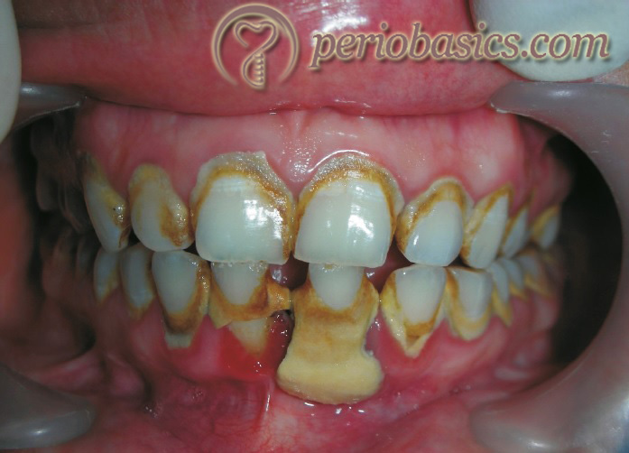
The calculus deposited on the teeth coronal to the gingival margin is designated as supragingival calculus. It is usually lighter in color (unless stained) and is less dense than subgingival calculus. Supragingival calculus almost always occurs predominantly on the lingual surface of the lower anterior teeth, with lesser amounts on the buccal surface of the upper molars. It has a hard clay-like consistency and can be easily detached from the tooth surface. It is also called as salivary calculus as most of its mineral component is derived from the saliva.
Subgingival Calculus
The calculus deposited on the tooth structure and found apical to the gingival margin within the periodontal pocket is designated as subgingival calculus. It is usually dense, dark brown or greenish-black in color, has a hard or flint-like consistency and it is firmly attached to the tooth surface 8. It is also known as seruminal calculus based on the assumption that most of the mineral content of subgingival calculus is derived from the gingival crevicular fluid 9, 10.
Differences between supra and subgingival calculus
| Features | Supragingival | Subgingival |
|---|---|---|
| Definition | Tightly adhering calcified deposit that forms on the crowns of the teeth above the free gingival margin. | Calcified deposit that forms on the tooth surface below the free margin of gingival and extends into the periodontal pocket. |
| Also known as | Salivary calculus. | Seruminal calculus. |
| Visibility | Visible in the oral cavity. | Below the crest of marginal gingival, usually in the periodontal pocket, not visible. |
| Location | Most abundant opposite the opening of major salivary glands, lingual surfaces of lower anteriors, buccal surfaces of upper first molars . | Usually seen in proximal surfaces of teeth and with less frequency on buccal surfaces |
| Prevalence | Usually common upto the age of 9 years 0-15 yrs: 37-70% 16-21 yrs: 44-88% Above 40 yrs: 86-100% | Slightly lower than that of supragingival, but approaches a range of 47- 100% after age of 40 yrs |
| Color | White or whitish yellow can be influenced with exposure to food, tobacco and with age. | Dark brown /green black. Not influenced by food and tobacco. |
| Consistency | Hard clay-like. | Dense flint-like. |
| Attachment | Lightly attached to the surface. | Firmly attached. |
| Source of minerals | Salivary secretions. | GCF |
| Plaque overlying | Composed of primarily filamentous organism oriented perpendicular to underlying calcified deposit. | Consist of cocci, rods, filaments with no distinct pattern of alignment. |
| Composition | Mainly brushite and octacalcium phosphate. | More amount of magnesium whitlokite, less of brushite and octacalcium phosphate. Higher concentration of Ca, Mg and F than supragingival calculus. |
| Salivary protein | Found. | Not found. |
| Sodium content | Decreased. | Increased. |
Composition and structure of dental calculus
Dental calculus is primarily composed of calcium phosphate mineral salts, deposited between and within the remnants of formerly viable microorganisms. The plaque formation serves as an organic matrix for the subsequent mineralization of the deposit. Calcification of dental plaque biofilm results from mineral ions provided by bathing saliva or crevicular fluid. The supersaturation of saliva and plaque fluid with calcium and phosphate ions is the driving force for plaque mineralization. Both salivary flow rate and plaque pH appear to……. Contents available in the book……………. Contents available in the book……………. Contents available in the book……………. Contents available in the book………
Organic components
The organic contents of supra and subgingival calculus contain amino acids, lipids, carbohydrates, and other macromolecules.
Amino acid content:
The most thorough analysis of the protein constituents of dental calculus was carried out by Osuoji and Rowles (1974) 12 when they investigated the amino acid content of demineralized acid hydrolysates of mixed supra- and subgingival calculus and subgingival calculus separately. Seventeen amino acids were detected with glutamic, aspartic, glycine, alanine, valine and leucine forming the largest proportion of the total residue isolated. The sulfur-containing amino acids, methionine, and cysteine were present only in trace amounts. Neither cysteine nor hydroxyproline was detected, suggesting the absence of keratin or collagen.
This information agrees closely with the earlier work of Little et al. (1966) 13 who additionally noted differences between the lower lingual and upper molar calculus samples where the amino acid concentration of the former was found to be lower and of a different composition. The nitrogen content of the upper molar calculus samples was also consistently higher than that of the lower anterior calculus.
Lipid Content:
The lipid content of dried calculus was studied by Osuoji and Rowles (1974) 12, using chloroform-methanol extraction. A combination of thin-layer chromatography and gas-liquid chromatography showed the presence of a variety of lipid components and fatty acids. The lipid content was 15.3% of the dry weight of the decalcified calculus and included phospholipids, cholesterol esters, diglycerides, triglycerides and free fatty acids. The free fatty acids represented the largest component of the lipid fraction and the predominant acids detected were palmitic, stearic and oleic, with smaller amounts of the unsaturated fatty acids, linoleic and linolenic acids.
Carbohydrate Content:
Studies by Little et al. (1966) 13 on acid hydrolysates of decalcified calculus indicated the presence of a variety of constituent sugars, including glucose, galactose, galactosamine, glucuronic acid, galacturonic acid, glucosamine, rhamnose and sometimes arabinose. No difference in carbohydrate content either chemically or chromatographically was found between different calculus sites or different mouths.
The constituent sugars yield some information on the source of origin of the calculus components. Hexuronic acid is a notable component of glycosaminoglycans present in connective tissue. It is not found in the saliva or in the cell wall of oral microorganisms. It is, however, a basic component of capsular Streptococcus and limes where it is present as hyaluronic acid and other unidentified uronides. The absence of deoxyribose and ribose preclude the presence of nucleic acids in calculus and indicates that the oral microorganisms undergo extensive degradation, leaving only the cell wall for calculus formation.
Macromolecules:
A variety of organic macromolecules have been identified in dental calculus. Embery (1977) 14 reported the presence of a sulfate glycopeptide in supragingival calculus, which contained a high amount of ester sulfate, hexose, equal amounts of galactosamine and glucosamine and a small quantity of peptide. In another study, Embery and Whitehead (1977) 15 noted the presence of sulfated glycosaminoglycans, hyaluronic acid in supragingival calculus and in addition to these sulfate glycosaminoglycans, chondroitin sulfate, and dermatan sulfate were also found to be present in subgingival calculus.
Inorganic components
Although, the process of mineralization is not completely understood, but it has been proposed to involve localized supersaturation, nucleation, crystal growth and the transformation of precursor phases such as dicalcium phosphate dihydrate, octacalcium phosphate, and amorphous calcium phosphate into more stable, crystalline deposits of hydroxyapatite and whitlockite 16. Supragingival and subgingival calculus contain 37% and 58% mineral content by volume, respectively 17. The principal inorganic elements present in calculus are 18,
- Calcium 39%
- Phosphorus 19%
- Carbon dioxide 1.9%
- Magnesium 0.8%
- Trace elements: Sodium, Zinc, Strontium, Bromine, Copper, Manganese, Tungsten, Gold, Aluminum, Silicon, Iron and Fluorine.
The crystalline structure of the inorganic content of supra and subgingival calculus consists of octacalcium phosphate [Ca8(PO4)4(HPO4)25HO], hydroxyapatite [Ca10(PO4)6(OH)2], and β-tricalcium phosphate or whitlockite [Ca10(HPO4) (PO4)6]. Brushite [CaHPO4-2H2O: dicalcium phosphate dihydrate] is present ……. Contents available in the book……………. Contents available in the book……………. Contents available in the book……………. Contents available in the book………
The organic matrix of dental calculus and its interaction with minerals
As already stated, the mechanisms by which the mineralization of dental calculus is initiated are not fully understood. In contrast to the molecular events which occur in the homeostasis of bone and dentine, dental calculus represents a surface phenomenon and is therefore outside the normal cellular, neural and hormonal controlling influences. As such there is no remodeling of the calculus and it increases in bulk via accretion due to surface activity. Limits on size will be imposed by shear forces in the mouth, particularly by fragmentation due to masticatory forces. Along with the calcification systems which occur in physiologic systems, the organic components present are considered to play an important role in the mineralization process. The major part of the organic matrix is derived from the saliva, gingival sulcus fluid, and oral bacteria, with a contribution from cell debris such as polymorphonuclear leukocytes.
In 1957 Mandel and Levy 23 suggested a role of protein-polysaccharide complexes in the formation of calculus. These workers demonstrated the presence of carbohydrate-protein molecules in supragingival calculus using histochemical procedures alone. Further studies by this group and by Little et al. (1966) 24 yielded valuable information on the carbohydrate and amino acid constituents of acid hydrolysates of the supra-gingival calculus from the molar and lingual areas of a large number of individual samples. Ennever et al. (1977) 25 contended that the initiator of calculus matrix calcification is proteolipid. The presence of glycosaminoglycan-rich proteoglycan metabolites in calculus are also potential nucleators due to their high number of sulfate and carboxyl groups which can attract calcium. These molecules, in addition to concentrating calcium ions, may attract water molecules and thereby locally increase the metastability of the calcium phosphate solution, leading to its deposition around the organic macromolecules.
These molecules can also inhibit or certainly regulate the calcium phosphate formation. Chen and Boskey (1984) 26 have shown that coating the hydroxyapatite seed crystals with proteoglycan, chondroitin sulfate, and dextran sulfate decreased the amount of hydroxyapatite precipitated as a function of time, whereas the desulfated analogues showed less inhibition. On the other hand, the removal of sulfate may induce conformational changes, indicating that charge may not be the only determinant during calculus formation. Many reviews have discussed in detail calculus formation and its prevention and a number of theories of calculus formation have been put forward. Some of the prominent theories regarding calculus formation have been described in the following discussion.
Theories of calculus formation
Mineralization theory:
According to this theory, saliva is saturated with various inorganic ions and thus it is able to support crystal growth by spontaneous precipitation of inorganic ions. However, this theory has a major drawback that spontaneous precipitation of inorganic ions does not occur unless the solution is seeded with nucleation centers for crystal growth.
Bacterial theory:
The importance of bacteria in the formation of calculus has historically received a considerable endorsement. Multiple mechanisms have been suggested by which bacteria may facilitate calculus formation. It has been proposed that bacteria may,
- Form phosphatases, which increase the local concentration of phosphates and thereby lead to calcification.
- Affect the pH of plaque and saliva and destroy protective colloidal action of the latter.
- Attach the calculus to the tooth.
- Provide chemicals that induce mineralization.
It has been shown that initial deposition of apatite in calcifying bacteria is associated with a membrane or acidic membrane-associated components 27. The ability of acid phospholipids in calculus formation depends on their ability to bind calcium by their negatively charged groups. Further-more, the calcifiability of bacteria is positively correlated with the increasing concentration of membrane-associated proteolipids 27.
An alternative view is that bacteria may only be passively involved in calculus formation and simply are calcified along with the other components of plaque. The occurrence of calculus formation in germ-free animals lends credence to such an opinion, but does not rule out active participation by bacteria had they been present. The viability of micro-organisms is not necessary for participation in mineralization since non-viable microorganisms calcify readily 28. Decrease in metabolic activity with reduced production of organic acids that result from glycolysis may be a pre-requisition before bacteria can mineralize.
Carbon dioxide theory:
This theory is based on the assumption that differences in the CO2 tension in saliva and the atmosphere may facilitate calculus formation. The freshly secreted saliva, leaving the opening of the salivary ducts has a CO2 tension of about of 60 mm Hg, in the expired air it is about 29 mm Hg and in the atmosphere, it is about 0.3 mm Hg. This discrepancy has been proposed to result in the escape of CO2 from the saliva causing rise in its pH. When the pH of saliva increase, less Ca and phosphorous can be accommodated in the ionized form and consequently, their spontaneous precipitation occurs. Once crystallites are formed, the physiological supersaturation results in their growth, resulting in calculus formation. This theory explains the formation of calculus near the orifices of the major salivary glands in a copious amount. However, it cannot explain the formation of subgingival calculus where the gingival crevicular fluid is the main source of calculus components.
Ammonia theory:
Ammonia is a breakdown product from urea and might result in local pH increase in plaque. The abundant supply of urea from the major salivary gland secretions tends to increase the pH in plaque 29. The increase in pH in plaque is primarily due to proteolytic activity in plaque which may result in the formation of amines, urea and ammonia 30. A positive correlation has been found between the proteolytic activity in plaque and calculus formation 31. Further, it has been demonstrated that the subgingival calculus index correlates well with the protease activity in salivary sediment, and the supragingival calculus index shows a significant correlation with the protease in salivary supernatant 32.
Booster concept:
According to this concept, one or more changes in the oral environment (booster mechanisms) may promote calculus formation, which include mechanisms facilitating an increase in pH of saliva, formation of supersaturated solutions of colloidal proteins in saliva and/or enzymatic mechanisms which facilitate the precipitation of calcium and phosphate ions. It has been demonstrated that ……. Contents available in the book……………. Contents available in the book……………. Contents available in the book……………. Contents available in the book………
Epitactic theory:
‘Epitaxy’ refers to crystal formation (e.g. hydroxyapatite) through seeding by another compound. According to this theory, seeding agents induce small foci of calcification that enlarge and coalesce to form a calcified mass 36. This concept has been referred to as the ‘epitactic concept’, or more appropriately, ‘heterogeneous nucleation’. The seeding agents in calculus formation are not known, but it is suspected that the intercellular matrix or plaque plays an active role 37. The carbohydrate-protein complexes may initiate calcification by removing calcium from the saliva (chelation) and binding with it to form nuclei that induce subsequent deposition of minerals. Plaque bacteria have also been implicated as possible seeding agents. The lipid component of the organic matrix has also been suspected as mineralization foci in plaque since in vitro experiments on the decalcified calculus reveal that extraction of the lipid component prevents remineralization of the calculus matrix 25.
Inhibition theory:
Another approach considers calcification as occurring at specific sites because of the existence of an inhibiting mechanism at non-calcifying sites. During calcification, the inhibitor is apparently altered or removed. One possible inhibiting substance is thought to be pyrophosphate (possibly other polyphosphates) and among the controlling mechanisms is the enzyme alkaline phosphate 38. The pyrophosphate inhibits calcification by preventing the initial nucleus from growing, possibly by “poisoning” the growth centers of the crystals.
Transformation theory:
The most attractive hypothesis is the idea that hydroxyapatite need not arise exclusively via epitaxy and nucleation. Amorphous ……. Contents available in the book……………. Contents available in the book……………. Contents available in the book……………. Contents available in the book……….
Rate of calculus formation and accumulation
The initiation, rate of calcification and accumulation of calculus vary from person to person, in different teeth, and at different times in the same person. On the basis of these differences, persons may be classified as:
1. Heavy calculus formers.
2. Moderate calculus formers.
3. Slight calculus formers.
4. Non-calculus formers.
The average daily increment in calculus formers is about 0.10% – 0.15% by weight 41, 42. Calculus formation continues until it reaches its maximum level, after which it may be reduced. The time required for calculus to reach its maximal level is variable from individual to individual and has been reported to vary from 10 weeks 43 to 6 months 44. The decline from maximal accumulation is referred to as “reversal phenomenon” which can be explained by the vulnerability of bulky calculus to mechanical wear from food and from the cheeks, lips and tongue.
Adhesional aspects of dental calculus formation
The adhesional bond strength of calculus to enamel and dentine surfaces determines the ease with which calculus can be removed by daily tooth brushing or professional dental treatment. Following mechanisms of attachment of calculus to the tooth surface have been proposed:
- Attachment by means of organic pellicle.
- Mechanical interlocking between the surface irregularities such as resorption lacunae and caries.
- Penetration of calculus bacteria into the cementum.
- A close adaptation of calculus under surface dispersions to the gently sloping mounds of the unaltered cementum surface.
Various studies have been done on animal and human models to evaluate the calculus attachment to different surfaces of the tooth. Beagle dog models have been extensively used for this purpose. It is important to emphasize at this point that two major differences exist between the beagle dog model employed and the human oral cavity regarding calculus formation. Firstly, calculus formed in beagle dogs is known to possess a high amount of calcium carbonates, whereas human calculus consistently contains calcium phosphates 45. Secondly, due to lack of regular brushing, the calculus surface in the dog model is rougher than generally observed in humans 46. This aspect may lead to increased rates of formation of calculus in dogs under the experimental conditions as compared to man.
The firmer adhesion of calculus to high surface free energy materials can be explained on the basis of the high surface free energies of both interacting surfaces, which is a thermodynamically favorable situation for adhesion. From a clinical point of view, it is important to realize that calculus removal can be facilitated by polishing the tooth surface thereby reducing its surface free energy. During instrumentation, easier calculus removal results in reduced loss of sound tooth structure. A major requirement for prevention of calculus formation is a smooth tooth surface. Once this is achieved, calculus formation can be reduced, but cannot be completely inhibited. As a second preventive measure, the tooth surface free energy may be reduced by adsorption of amine fluoride or perfluoroalkyl surfactants to facilitate easier removal of calculus 47.
Inhibitors of calculus formation
Calcification nucleation inhibitors:
One of the earliest identified calcification nucleation inhibitors is magnesium (Mg). Mg blocks apatite crystallization and stabilizes calcium phosphate as amorphous mineral 48. Other nucleation inhibitors include diphosphonates which inhibit both apatite nucleation 49 and crystal growth 50. These agents, however, are not used clinically because they interfere with the normal process of hard tissue mineralization.
Crystal growth inhibitors in saliva:
Various salivary proteins have been investigated for their inhibitory action on crystal growth. These proteins get adsorbed at active sites on the crystalline surfaces, thus inhibiting the growth and dissolution of calcium phosphate crystals. The most extensively studied of these proteins are proline-rich proteins (PRPs), statherin, and cystatins. The inhibition of crystal growth by PRPs has been studied in an in vitro study by Margolis et al. (1982) 51. In this study, it was demonstrated that proline-rich protein-3 reduced the crystallization rate, but did not completely block the crystal growth. In another study, Bennick et al. (1979) 52 investigated the adsorption sites of PRPs. They found that ……. Contents available in the book……………. Contents available in the book……………. Contents available in the book……………. Contents available in the book………
Promoters of calculus formation
Urea:
One of the most common promoters of calcification is urea. It is the product of the metabolism of nitrogen-containing compounds. Urea metabolism is closely associated with plaque mineralization. The concentration of urea in saliva varies from 5 to 10 mmol/L 54. However, in patients with renal failure, its concentration in saliva has been found to be as high as 30 mmol/L 55. The ureolysis of urea leads to the formation of ammonia, which contributes in increasing the pH of plaque which promotes calculus formation by increasing the degree of saturation of calcium phosphate in the plaque fluid. The urea dependent pH response in plaque is affected by the salivary flow. Urea in the oral cavity is derived from the saliva and extra-oral factors such as urea-containing chewing gums and toothpaste. In the absence of exogenous urea, the increased salivary flow rate promotes the urea-dependent pH response in plaque. Thus, increased salivary flow sites are associated with an increased rate of calculus formation. This factor partially explains the high rate of calculus formation at particular sites in the dentition 56.
Ureolysis by bacteria in dental plaque has also been proposed to play an important role in the calculus formation. Bacteria in plaque which have been shown to have ureolytic activity include Streptococcus salivarius, coagulase-negative Staphylococci 57,58, Actinomyces viscosus/naeslundii 59, transient Enterobacteriaceae, unknown anaerobes 60, and Haemophilus species 61. The most investigated bacteria out of the above is S. salivarius which is a minor component of plaque, and has been shown to play an important role in plaque ureolysis 62.
Fluorides:
Fluorides have been well known for their anti-caries activity. The substitution of hydroxyl ions by fluoride ions in hydroxyapatite crystals results in the formation of fluorhydroxyapatite, which has low solubility 63. Fluorides also facilitate remineralization by precipitation of a fluoride-enriched apatite or calcium fluoride 64. Furthermore, fluorides have been shown to affect the morphology of the apatite crystals, converting them from thin plates to short and slender needles which further reduces their solubility 65.
Fluorides also have been shown to have antibacterial action 66 and to inhibit acid production by the bacteria 67. Thus, fluorides may have the potential to increase the plaque pH, which may contribute to increased calculus formation. Thus, it can be assumed that fluorides present in the toothpastes and mouthwashes may increase the calculus formation. However, there is only scanty evidence in support of this assumption. On the contrary, it has been shown that sodium fluoride may be able to inhibit the bacterial enzymes namely, phosphatases and pyrophosphatases, which are known to increase calculus formation 68.
Silicon:
Silicon is found in drinking water and food, commonly in the form of silicic acid and silica. The silicic acid family consists of mono- and poly salicylic acids which have been shown to be strong promoters of spontaneous precipitation of calcium phosphates 69. Silica and silicic acids have the calcium-binding capacity and they stimulate calcium phosphate precipitation. Thus, they can be considered as potent promoters of calculus formation. A study done on Indonesian and Norwegian population demonstrated that rate of calculus formation was more in the Indonesian population which was accounted for the consumption of larger amounts of rice by the Indonesian population, which is enriched in silicon 70.
Detection of calculus
Detection of calculus and its complete removal is mandatory to achieve a clean tooth surface. It is the first and mandatory step in periodontal therapy. The clinician should be well trained to effectively remove calculus from the tooth surface, but at the same time prevent any damage to the tooth surface.
Conventional calculus detection techniques:
The most commonly used method for the detection of calculus is ‘Direct vision’. But it is limited for the detection of supra-gingival calculus only. Compressed air may be put on the gingival crevice to displace the gingival margin, thus facilitating the viewing of subgingival calculus. But still, it may not allow the visualization of the deeper calculus.
Another commonly used technique for the detection of calculus is ‘tactile sense of the operator‘. For this, the operator uses a probe, explorer or a curette to detect the presence of subgingival calculus. The end result of scaling and root planing should be a smooth root surface. Studies have shown that excess amount of root surface is removed by clinicians to achieve a smooth root surface in case of subgingival scaling and root planing 71.
One more, commonly used detection technique for the detection of calculus is ‘radiographic technique’. In this calculus can be seen on a radiograph as a radiopaque discontinuity on the root surface, but this technique is only occasionally useful where an adequate mass of deposit is present on the root surface. This technique is not useful to detect the presence of residual calculus on the root surface after scaling and root planing.
To overcome the limitations of conventional calculus detection techniques, many recent techniques have been introduced which are able to detect and /or remove the subgingival calculus from the tooth surfaces.
Advancements in calculus detection techniques:
Following is the brief description of various calculus detection and calculus detection and removal systems,
PERIOSCOPY™ (Perioscopy Inc., Oakland, California)
Perioscopy system is a modification of medical endoscope. It is exclusively used for periodontal purposes. It was developed in the year 2000. It consists of a fiber-optic bundle surrounded by multiple illumination fibers, a light source, and an irrigation system. Its miniature design causes minimal tissue trauma. The fiber-optic system permits visualization of the subgingival root surface and calculus deposits in real-time on a display monitor 71.
DetecTar™ (Ultradent Inc. South Jordan, Utah)
DetecTar™ uses a spectro-optical approach in order to detect subgingival calculus by utilizing a light-emitting diode and fiber optic technology. This involves an optical fiber which recognizes the characteristic spectral signature of calculus caused by absorption, reflection, and diffraction of red light 71.
DIAGNODENT™ (KaVo Dental, Charlotte, NC)
This system is based on the fact that the composition and structure of calculus and tooth surface are different. This structural difference gives a typical fluorescence to both these structures. Calculus contains various ……. Contents available in the book……………. Contents available in the book……………. Contents available in the book……………. Contents available in the book………
Keylaser3™ (KaVo Dental, Charlotte, NC)
It is a laser system that combines a 655nm InGaAsP diode for detection of calculus and a 2940 nm Er: YAG laser for treatment. Previous versions of this system (i.e. Keylaser 1 and 2) can be used for the removal of calculus only. A scale of 0-99 is used for the detection of calculus. Values exceeding 40 indicate the definite presence of mineralized deposits. Er: YAG laser is activated as a certain threshold is reached. As soon as the value falls below the threshold level, Er: YAG laser is switched off. Studies done to assess the efficacy of this device have shown that it produces tooth surface comparable to hand and ultrasonic instruments. Cost factor can be a limiting aspect of using lasers for detection and treatment 71.
PERIOSCAN™ (Sirona, Germany)
It is an ultrasonic device that works on acoustic principles. It is similar to tapping on a glass surface with a hard substance and analyzing the sound produced to find out the cracks that are present on the glass. Tip of the ultrasonic insert is oscillating continuously. Different voltages are produced due to changes in oscillations depending on the hardness of the surface. The hardness of the calculus differs from the hardness of the tooth surface. This difference in hardness can be used to generate the information of the surface that is being touched by the device. It can differentiate between calculus and healthy root surfaces. It also has a treatment option that can be used to remove these calculus deposits immediately. This combination of detection and removal mechanism is advantageous since calculus can be removed just by switching the mode from detection to removal. The advantage lies in the fact that relocating the previously located calculus is not necessary 71.
Pathogenic potential of dental calculus
Until 1960, the prevailing view was that the calculus is a major etiologic factor in periodontal disease. Its pathogenicity was attributed to its rough outer surface, which mechanically irritated the adjacent tissues. Shroeder (1969) 72, stated that the initial damage to the gingival margin in periodontal disease is due to immunologic and enzymatic effects of the microorganisms in plaque. However, the process is enhanced by the presence of supra- and subgingival calculus, which provides surface for new plaque accumulation. It is still not clear, however, whether calculus plus plaque provokes a greater reaction than plaque alone, although there is some suggestive evidence for the former 73. There is no question, however, that the mineralized deposits do,
- Bring the bacterial overlay closer to the supporting tissues.
- Interfere with local self-cleansing mechanisms.
- Make plaque removal more difficult for the patient.
It is difficult to separate the effects of calculus and plaque on the gingiva because calculus is always covered with a non-mineralized layer of plaque. There is always a positive correlation between the presence calculus and prevalence of gingivitis, but this correlation is not as great as that between plaque and gingivitis. Longitudinal and cross-sectional epidemiological studies have demonstrated clear associations between the presence of calculus and periodontitis 74-81. However, most of the studies have not demonstrated a direct cause and effect relationship between calculus and periodontal disease progression. However, regardless of its primary or secondary relationship in pocket formation, and although the principal irritating features of calculus is its surface plaque rather than its calcified interior, calculus is a significant pathogenic factor in periodontal disease.
Subgingival calculus because of its presence below the gingival margins is generally considered more deleterious as compared to supragingival calculus. A number of studies support the view that the presence of subgingival calculus contributes to the chronicity and progression of periodontal diseases 73. Clinical studies attest to the importance of frequent and thorough removal of root deposits by scaling and root planing to prevent attachment loss 82. Morphological studies show that calcified deposits are porous and could act as a reservoir for irritating substances. Experimental studies have established the permeability of subgingival calculus to endotoxin 83 and the presence of high levels of toxic stimulators of bone resorption and antigens from Porphyromonas gingivalis in the calculus 84.
Thus, from the above discussion, it is clear that a thorough removal of porous bacterial retentive subgingival calculus is a key phase in periodontal therapy. Partial inhibition of plaque mineralization can be accomplished by chemical agents, but there has been no demonstration of a reduction of gingivitis in humans 85.
Anti-calculus agents used in commercial dentifrices
Triclosan with a PVM/MA copolymer
Triclosan [5-chloro-2-(2,4-dichlorophenoxy)phenol] is a broad-spectrum antibacterial agent which is active against both Gram-positive and Gram-negative microorganisms. The bacteriostatic activity of triclosan is attributed to prevention of essential amino acid uptake by the bacteria, while bactericidal activity is attributed to the disruption of the integrity of the cytoplasmic membrane, resulting in leakage of cellular contents 86. It is also a potent inhibitor of both cyclooxygenase and lipoxygenase pathways 87. The anti-bacterial activity of triclosan is ……. Contents available in the book……………. Contents available in the book……………. Contents available in the book……………. Contents available in the book………
Pyrophosphate and PVM/MA copolymer
Pyrophosphate is a small molecule that has been reported to inhibit crystal growth by binding to the surface of the crystal. There are two sites on the hydroxyapatite crystal surface where Pyrophosphate molecules bind. One of these two sites need to be bound by phosphate ion to permit crystal growth to occur. Attachment of pyrophosphate molecule at this site blocks adsorption of phosphate ion onto the crystal, and thus crystal growth is inhibited. To inhibit crystal growth effectively, the concentration of pyrophosphate has to reach a critical level. Below this level, the addition of NaF can induce a short period of slow precipitation, which is followed by rapid crystal growth 92.
Zinc ions
Zinc is added to the toothpastes and mouth rinses, as an anti-bacterial agent to help control plaque, to reduce oral malodor and to reduce calculus formation through crystal growth modification/inhibition 93-95. Zinc ions have been found to reduce the acidogenicity of plaque and inhibit its formation 96. There is evidence that zinc ions may inhibit both the adso-rption of bacteria to the tooth surface and growth of existing plaque 97, 98. Zinc competes with calcium for bacterial binding sites which imply that it reduces plaque formation. Zinc has a good oral substantivity. Following application, relatively large amounts of the applied zinc dose are retained in the mouth, with reported values typically between 15-40% 99-103.
Conclusion
In the present chapter, we studied various aspects of dental calculus including its biochemical and structural organization. Calculus provides an ideal surface for plaque accumulation and can be therefore considered as a secondary etiological factor in the development of periodontitis. Its removal from the tooth surface is a primary requirement to achieve periodontal health. The clinician should be well trained in the adequate removal of calculus, which is the first step in periodontal therapy. The design and sharpness of instruments, anatomical factors, depth of calculus deposition and operator’s experience all play an important role during subgingival calculus removal. Although, many new techniques and equipment have been introduced which can detect and /or remove calculus, but these have not shown much benefit as compared to conventional ultrasonic devices. Hence, one may use any individual or a combination of techniques, to achieve the ultimate goal of calculus removal.
References
References available in the hard copy of the website.