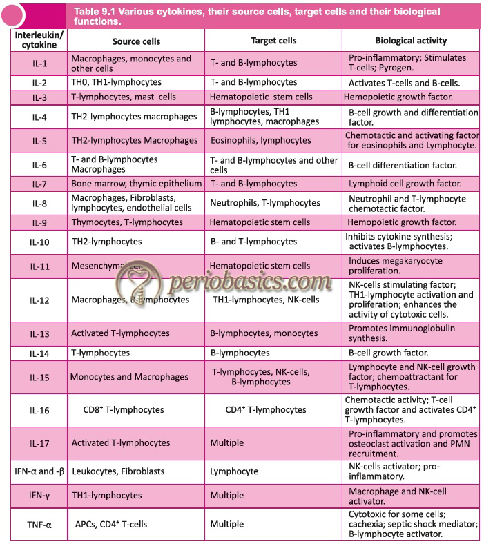
Introduction to cytokines
Cytokines (in Greek ”Cyto-”, cell; and ”-Kinos”, movement) are the substances that are secreted by specific cells of the immune system which carry signals locally between the cells, and thus have an effect on other cells. A special subset of cytokines is the interleukins (ILs), of which 23 different types have been characterized. The biological effects of cytokines are extremely diverse as they influence not only the immune response but also inflammatory processes and hematopoiesis. They can be classified into the following categories:
- Pro-inflammatory cytokines.
- Cytokines with predominant immunoregulatory functions.
- Cytokines that regulate lymphocyte growth, activation, and differentiation.
- Cytokines that help in hematopoiesis.
- Chemokines.
In the following sections, these cytokines have been discussed in detail.
Pro-inflammatory Cytokines
These are the cytokines that mediate natural immunity. In this group, there are soluble factors that influence inflammatory reactions. These include IL-1, tumor necrosis factor (TNF), type I interferons, IL-6, IL-8, and macrophage migration inhibition factor (MIF).
Interleukin-1 (IL-1):
This cytokine has diverse activities and roles in immunity, inflammation, tissue breakdown, and tissue homeostasis 1-4. IL-1 was originally discovered as a factor that induces fever. Later, its two molecular forms, IL-1α and IL-1β, encoded by two separate genes and displaying only 20% homology to one another, were identified. Cells of the monocyte-macrophage lineage are the main cellular source of IL-1, though most cell types have the potential to express this cytokine. IL-1 synthesis is primarily in response to bacterial derived endotoxins. IL-1α tends to remain associated with the cell membrane, while IL-1β (synthesized as an inactive precursor) is released from the cell after being processed post-translationally by a cysteine-asparagine protease.
IL-1β is a pro-inflammatory cytokine expressed by monocytes, macrophages, and dendritic cells. It signals through two receptors, IL-1RI and IL-1RII, both of which are shared with IL-1α. It is a multifunctional molecule with effects ranging from inflammation, immunity, and hemopoiesis.
Tumor necrosis factor (TNF):
Tumor necrosis factor exists in two forms TNF-α and TNF-β.
TNF-α:
TNF-α is produced by various cells and is involved in normal inflammatory and immune responses. It is both autocrine and paracrine inducer of other cytokines like, IL-1, IL-6, IL-8, platelet-derived growth factor-β, eicosanoids, platelet-activating factors and granulocyte monocyte colony-stimulating factor. TNF-α is secreted by macrophages, monocytes, neutrophils, T-cells, Natural Killer cells (NK-cells) following their stimulation by bacterial lipopolysaccharides. Cells expressing CD4 secrete …………Contents available in the book………….Contents available in the book………….Contents available in the book………….Contents available in the book….

Periobasics: A Textbook of Periodontics and Implantology
The book is usually delivered within one week anywhere in India and within three weeks anywhere throughout the world.
India Users:
International Users:
TNF-β:
TNF-β is commonly known as lymphotoxin. In humans, it is encoded by the LTA gene. It is a potent multifunctional cytokine produced mainly by activated T- and B-lymphocytes. This cytokine has been shown to have a specific potent cytotoxic activity on tumor cells and a variety of other target cells. It has also been shown as a potent mediator of inflammation and immune function.
IL-1 and TNF have effects at three different levels 9:
Metabolic effects:
IL-1 and TNF-α induce the synthesis of acute-phase reactants. These include many proteins such as α1-antitrypsin, fibrinogen, and C-reactive protein in the liver. These are increased in situations associated with inflammatory reactions.
Vascular effects:
IL-1 profoundly affects the function of vessel wall elements, in particular, that of endothelial cells. IL-1 activates endothelial cells in a pro-inflammatory, pro-thrombotic sense. IL-1 induces the production of tissue factor and platelet-activating factor. IL-1α and TNF-α promote the adherence of inflammatory cells, which eventually migrate into the extravascular space, where they form tissue inflammatory infiltrates. They particularly promote the expression of P-selectin and E-selectin, in vascular endothelial cells.
Endogenous pyrogen:
TNF-α is a potent endogenous pyrogen and acts on cells in hypothalamic regulatory regions of the brain to induce fever. Although IL-1 does not cross the blood-brain barrier, but it acts on the periventricular organs where the blood-brain barrier is interrupted. It interacts with a group of nuclei in the anterior hypothalamus, causing fever.
Know More…
Properties of cytokines:
Pleiotropy:
A cytokine is said to have pleiotropic property when it has different biological effects on different target cells.
Redundancy:
Two or more cytokines that mediate similar functions are said to be redundant. Redundancy makes it difficult to ascribe a particular activity to a single cytokine.
Synergy:
When the combined effect of two cytokines on the cellular activity is greater than the additive effects of the individual cytokine, then it is referred to as synergism.
Antagonism:
When the effects of one cytokine inhibit or offset the effects of another cytokine, it is referred to as antagonism.
Cascade induction:
Cascade induction occurs when the action of one cytokine on a target cell induces that cell to produce one or more other cytokines, which in turn may induce other target cells to produce other cytokines.
Type-I interferons
The type-I interferons (IFN) i.e., IFN-α and IFN-β have pro-inflammatory action along with their predominant immuno-regulatory functions (discussed later).
Interleukin-6 (IL-6)
IL-6 along with IL-1 and TNF is a major immune and inflammatory mediator. It is a pleiotropic cytokine influencing immune responses and inflammatory reactions 10. It is produced by a variety of cells, such as monocytes, fibroblasts 11, osteoblasts 12, and vascular endothelial cells 13 in response to inflammatory challenges 14. It has pro-inflammatory and hematopoietic activities. Its role as a pro-inflammatory cytokine is similar to that of IL-1 and TNF, particularly in the induction of the synthesis of acute-phase response proteins by the liver. One action of IL-6 on B-cells is the increased secretion of IgM 15. It also induces T-cell proliferation 16. It acts along with IL-2 to induce T-cell proliferation and cytolytic effector cell generation.
Interleukin-8 (IL-8)
In humans, IL-8 is one of the fifteen members of the CXC chemokine family. It is known for its effects on neutrophils, especially its ability to act as a chemoattractant for them 17-19. In addition to its effects on neutrophils, IL-8 promotes angiogenesis, inhibits endothelial cell apoptosis, and promotes the proliferation of melanoma cells in an autocrine fashion 17. This cytokine is induced and secreted by many cells, such as monocytes 20, lymphocytes 21, fibroblasts 22, epithelial and endothelial cells 23, 24 as well as by synovial cells 22. It induces the adhesion of PMNs to endothelial cells and their transendothelial migration 25 as well as the release of granule enzymes from these cells 26.
Macrophage migration inhibition factor (MIF)
MIF plays crucial roles in the recruitment and activation of macrophages as well as in helping them to kill bacteria. It is also known as a glycosylation-inhibiting factor (GIF). MIF plays a major role in innate immunity against bacterial infections through enhancement of tumor necrosis factor (TNF)-α secretion 27, Toll-like receptor 4 (TLR4) expression 28, phagocytosis and intracellular killing mechanisms 29 and is equally efficiently involved in the adaptive immune response by favoring Th1 activation and differentiation 30, 31.
Cytokines with predominant immunoregulatory functions
In this category, we have interferons, IL-12 and IL-18.
Interferons (IFNs)
The IFNs are a large family of multifunctional secreted proteins involved in antiviral defense, cell growth regulation, and immune activation. They amplify the antigen presentation to T-cells and also increase the resistance of uninfected cells to viral infection. Furthermore, they are also involved in the activation of macrophages and B-cells. They are primarily involved in various immune interactions, acting as inducers, regulators, and affect both innate and acquired immune responses. Interferons present in humans are mainly classified into three types: α-interferons, β-interferons, and γ interferons. α interferons and β interferons are the members of Type I interferons whereas γ-interferon is a member of Type II interferons.
α-interferons:
They are primarily involved in innate immune response against viral infections. It is a large family of interferons containing around 13 subtypes, including IFNα-1, IFNα-2, IFNα-4, IFNα-5, IFNα-6, IFNα-7, IFNα-8, IFNα-10, IFNα-13, IFNα-14, IFNα-16, IFNα-17 and IFNα-21. These are predominantly synthesized by leukocytes.
β-interferons:
They are also involved in the innate immune response. These are further divided into three subtypes IFNβ-1, IFNβ-2, and IFNβ-3. These are synthesized by most cell types, but particularly by fibroblasts. IFNβ-1 is used in the treatment of multiple sclerosis.
γ-interferons:
There is only one type of IFN-γ. The human IFN-γ protein is a 166 amino acid polypeptide. It has a wide range of effects. In concert with IL-1β, it induces an increase in the expression of ICAM-1 on the cytoplasmic membrane of endothelial cells, enhancing T-lymphocyte adherence to the vascular endothelium, an essential step for T-lymphocyte egress from the vascular bed. Large numbers of T-lymphocytes will thus exit the vascular bed in areas near the tissues where activated T-cells are releasing interleukins and will form perivenular infiltrates characteristic of delayed hypersensitivity reactions. The most important effect of IFN-γ seems to be the activation of monocytes. After exposure to IFN-γ, monocytes undergo a series of changes typical of their differentiation into phagocytic effector cells:
- The cell membrane becomes ruffled; the number of cytoplasmic microvilli increases by a factor of 10. This change reflects a considerable increase in phagocytic capacities.
- The expression of MHC Class II antigens and Fc receptors increases. The increase in the expression of MHC Class II enhances the efficiency of monocytes as antigen-presenting cells (APCs), and the increased expression of Fc receptors further enhances their efficiency as phagocytic cells.
- The production of cytokines (TNF, IL-12) and of several antimicrobial proteins and compounds [including defensins, cathepsins, collagenases, superoxide radicals, and inducible nitric oxide synthase (iNOS)] is upregulated. As a consequence, engulfed organisms are rapidly killed and ingested proteins are efficiently digested in the phagolysosomes. It must be noted that excessive and protracted production of IFN-γ may have adverse effects. Hyper-stimulated monocytes may become exceedingly cytotoxic and may mediate tissue damage in inflammatory reactions and autoimmune diseases.
Another important function of IFNs is …………Contents available in the book………….Contents available in the book………….Contents available in the book………….Contents available in the book….
Interleukin-12 (IL-12)
IL-12 is an important regulatory cytokine that has a central function in the initiation and regulation of cellular immune responses. IL-12 is produced by macrophages, monocytes, dendritic cells, and B-cells in response to bacterial products and intracellular parasites. The biological effects of the production of IL-12 are directed at T-cells and NK-cells. IL-12 is primarily responsible for the subsequent production of IFN-γ and TNF-α from both NK-cells and helper T-cells.
It is produced by APCs after endocytosis of particulate materials or microbes and plays a crucial role in linking innate and adaptive immunity. IL-12 induces the production of IFN-γ by CD4 T-cells and NK cells in the early stages of the immune response, before the differentiation of effector T-cells.
Interleukin-18 (IL-18)
This interleukin was initially named as an interferon-γ inducing factor, reflecting its major biological role. However, it is a member of the IL-1 family that promotes the production of various pro-inflammatory mediators. It is synthesized and secreted by various cells, including macrophages, dendritic cells, intestinal epithelial cells, synovial fibroblasts, keratinocytes, Kupffer cells, microglial cells, and osteoblasts. Its main target cells are TH0/TH1 CD4 T-cells and NK-cells. It promotes T- and NK-cell maturation and also promotes secretion of TNF-α, IFN-γ, and GM-CSF. Other functions of IL-18 include increased production IL-4, IL-10, and IL-13, increased IgE expression on B-cells and in association with IL-2, an enhanced stimulus-induced IL-4 production from TH2 cells.
Cytokines that regulate lymphocyte growth, activation, and differentiation
This category of cytokines includes IL-2, IL-4, IL-5, IL-6, IL-12, IL-15 and transforming growth factor-β (TGF-β).
Interleukin-2 (IL-2)
IL-2 is a secreted cytokine that is important for the proliferation of T- and B-lymphocytes. Its structure consists of a single polypeptide chain of 133 amino acid residues. When a helper T-cell binds to an antigen-presenting cell using CD28 and B7, CD4 cells produce IL-2. This cytokine is necessary for the proliferation and differentiation of T-cells. During initial maturation, the unstimulated (resting) T-cells belonging to either the CD4 or the CD8 subsets possess a few high-affinity IL-2 receptors, but upon stimulation (by antigen), the number of IL-2 receptors increases significantly on their surface. The receptor of this cytokine is a heterotrimeric protein complex whose gamma chain is also shared by IL-4 and IL-7.
Interleukin-4 (IL-4)
IL-4 is a pleiotropic cytokine produced by TH2 cells, mast cells, and NK-cells. It is an important factor affecting both T- and B-cells. The primary function of IL-4 is the regulation of the differentiation of antigen-activated naïve T-cells.
Effect on T-cells:
It plays a major role in T-cell development. It is thought to be influential in promoting the differentiation of T-helper cells into TH2 cells during an immune response. IL-4 suppresses the production of TH1 cells. IL-4 can also act as a mast cell growth factor. IL-4 actions (except increases in MHC Class II) are “neutralized” by IFN-γ, which is made by TH1 cells.
Effect on B-cells:
IL-4 exerts different effects on B-cells at different stages in the cell cycle. On resting B-cells, IL-4 acts as an activating factor, inducing them to enlarge in size and increase Class II MHC expression. Following activation by an antigen or mitogen, IL-4 acts as a growth factor, driving DNA replication in the B-cells. In the case of proliferating B-cells, IL-4 acts as a differentiation factor by regulating class switch to C-epsilon and C-gamma-1, i.e., the production of the IgE and IgG1 subclasses. In this role, it has been termed a “switch-inducing” factor.
Interleukin-5 (IL-5)
IL-5 is secreted predominantly by TH2 lymphocytes. It can also be found in mast cells and eosinophils. IL-5 is best known for its activity on B-cells and eosinophils. Relative to B-cells, IL-5 appears to induce the differentiation of activated conventional B2 B-cells into Ig-secreting cells. IL-5 exerts its biological effects by binding to its membrane-bound receptor, IL-5R.
Interleukin-6 (IL-6)
It is a pro-inflammatory cytokine and is also known as B-cell differentiation factor. It is synthesized by mononuclear phagocytes, vascular endothelial cells, fibroblasts and other cells. The release of IL-6 by macrophages is triggered by …………Contents available in the book………….Contents available in the book………….Contents available in the book………….Contents available in the book….
Periobasics: A Textbook of Periodontics and Implantology
The book is usually delivered within one week anywhere in India and within three weeks anywhere throughout the world.
India Users:
International Users:
Interleukin-12 (IL-12)
It has the capacity to regulate the differentiation of naïve T-cells into TH1-cells. IL-12 is produced by macrophages, monocytes, dendritic cells, and B-cells in response to bacterial products and intracellular parasites. The biological effects of the production of IL-12 are directed at T-cells and NK-cells. IL-12 is primarily responsible for the subsequent production of IFN-γ and tumor necrosis factor-α (TNF-α) from both NK-cells and helper T-cells.
Interleukin-15 (IL-15)
IL-15 regulates T- and NK-cell activation and proliferation. This cytokine and IL-2 share many biological activities. They are found to bind common hematopoietin receptor subunits, and may compete for the same receptor, and thus negatively regulate each other’s activity. The number of CD8 memory cells is shown to be controlled by a balance between this cytokine and IL-2.
Details about transforming growth factor-β are available in “Application of growth factors in periodontal regeneration”.
Cytokines that help in hematopoiesis
Many cytokines stimulate the production of new blood cells by acting on hematopoietic progenitor cells. These include,
Granulocyte-monocyte colony-stimulating factor (GM-CSF)
It is one of the major cytokine stimulating hematopoiesis. It is released by activated T-lymphocytes. Its release stimulates granulocyte and monocyte production and their release from the bone marrow. It also stimulates monocyte cytotoxic activity and expression of MHC Class II molecules, dendritic cell proliferation and differentiation, and B-cell proliferation and differentiation.
Interleukin-1 (IL-1)
It basically leads to the mobilization of leukocytes which is objectively reflected in peripheral blood as leukocytosis. It acts as a growth factor in the bone marrow.
Interleukin-3 (IL-3) and stem cell factor
They are released mainly by activated T-lymphocytes and are important growth factors for early bone marrow progenitors, inducing the production of a wide variety of leukocytes.
Interleukin-7 (IL-7)
This interleukin is predominately produced by immune cells, such as T-cells, NK-cells, monocytes, and stromal or non-hematopoietic cells. It has pleiotropic immune regulatory functions and its main role seems to be the stimulation of the proliferation of B- and T-lymphocyte precursors.
Chemokines
Cytokines that regulate the leukocyte movements are also known as chemokines. In the chemokines, one amino acid separates the first two cysteine residues (Cys-X amino acid- Cys) and for that reason are also known as CXC chemokines. Chemokines have been classified into four main …………Contents available in the book………….Contents available in the book………….Contents available in the book………….Contents available in the book….
As discussed previously, IL-8 (neutrophil-activating factor) is the most important of all the chemokines. It is released by T-lymphocytes and monocytes stimulated with TNF or IL-1. It is also secreted by fibroblasts and endothelial cells. It functions as a chemotactic and activating factor for granulocytes, the cell population with the highest level of IL-8 receptor expression. IL-8 recruits granulocytes to areas of inflammation and increases their phagocytic and pro-inflammatory abilities. It has also been demonstrated to be chemotactic for T-lymphocytes.
Other chemokines include,
- RANTES (regulated on activation, normal T-cell express-ed and secreted) released by T-cells which attracts T-cells with memory phenotype,
- NK-cells, eosinophils, and mast cells.
- Macrophage inflammatory proteins (MIP), released by monocytes and macrophages that attract eosinophils, lymphocytes, and NK-cells.
- Macrophage chemotactic proteins (MCP), produced by monocytes, macrophages, and related cells, which attract monocytes, eosinophils, and
- NK and LAK (Lymphokine activated killer) cells.
- Eotaxin, a chemokine induced by IL-4 that recruits eosinophils and TH2 CD41 T-cells to the sites of allergic inflammation.
Role of cytokines in periodontal diseases
Various studies have shown that IL-1α, IL-1β, and TNF- α stimulate bone resorption and inhibit bone formation 33-35. Research work has shown that IL-1β expression is elevated in gingival crevicular fluid at sites of recent bone and attachment loss in patients with periodontitis 36. Similarly, studies done on non-human primate models using IL-1 antagonists {IL-1 receptor type I (IL-1R)}have shown a significant reduction in inflammation, connective tissue attachment loss, and bone resorption induced by periodontal pathogens as compared to controls 37. In another study, it was shown that the exogenous application of recombinant human IL-1β in a rat ligature model accelerated alveolar bone destruction and inflammation over a 2-week period 38.
Studies done on TNF-α have shown that it acts as a potent stimulator of bone resorption. The effect of recombinant human TNF- α (rhTNF- α) on the inflammatory response and periodontal breakdown in a rat ligature model was investigated in one study. Results showed the accelerated progression of periodontitis in rats 39. Similarly, TNF-α receptor-1-knockout (TNFR-1–KO) mice developed significantly less inflammation and alveolar bone loss in response to Aggregatibacter actinomycetemcomitans oral gavage in one study 6.
IL-4 has anti-inflammatory properties mediated by …………Contents available in the book………….Contents available in the book………….Contents available in the book………….Contents available in the book….
Further, it has been shown that IL-4 is also able to inhibit the production of matrix metalloproteinases (MMPs) and RANKL, and concomitantly inducing the upregulation of their respective inhibitors TIMPs and OPG 48, reinforcing its potential protective role in periodontal disease pathogenesis 49.
IL-6 is a potent pro-inflammatory cytokine. Investigations on IL-6 have reported that IL-6 levels in inflamed gingival tissues were higher than those in healthy control tissues 11. Studies done on mice with genetically deleted IL-6 had decreased bone loss as compared to wild-type mice in response to P. gingivalis, suggesting that the production of IL-6, which is pro-inflammatory, contributed to bone resorption 50. One of the major functions of IL-6 is the induction of the final maturation of B-cells into immunoglobulin-secreting plasma cells. It stimulates the secretion of antibodies to such a degree that serum IgG1 levels can rise up to 120-400-fold. In inflammatory periodontal lesions, a variety of cell types such as T-cells, macrophages, endothelial cells, and fibroblasts have been shown to express IL-6 at both mRNA and protein levels 51-54. IL-1β and IL-6 have also been characteristically associated with inflammatory cell migration and osteoclastogenesis processes 7, 55.
As already explained, IL-8 is a potent chemotactic factor for leukocytes 56, 57. It is secreted by a variety of cells, including monocytes, fibroblasts, lymphocytes, and endothelial cells. IL-8 induces neutrophil extravasation at the site of inflammation. One study found that IL-8 concentrations were significantly higher in gingiva adjacent to probing pocket depth ≤3 mm and less adjacent to > 6 mm sulci 58. Some other investigations suggested the absence of a direct relationship between IL-8 and PMN recruitment 59 and no significant difference in GCF IL-8 levels between localized juvenile periodontitis and healthy subjects 60.
Studies have shown anti-inflammatory activity of IL-11 61. This interleukin has been shown to play an important role in the modulation of the immune response via the reduction of pro-inflammatory cytokine production in animal models 61. It was predicted that IL-11 may be considered as a candidate molecule for therapeutic modulation of the host response in the management of periodontal diseases 62, 63. The anti-inflammatory action of IL-10 has also been demonstrated in one study. In this study, it has been suggested that a large number of infiltrating inflammatory cells, as well as accessory cells, are involved in the down-regulation of the inflammatory and immune response in periodontitis by releasing anti-inflammatory cytokines 64.
Know More…
Anti-cytokine therapy:
The anti-cytokine therapy targets the pro-inflammatory cytokines, including TNF-α, IL-1, and IL-6, etc., which are involved in the initiation of periodontal diseases. The anti-cytokine agents exert their effect in three ways,
By neutralization of cytokines,
By blockage of cytokine receptors, and
By activation of anti-inflammatory pathways, such as immune-suppressive pathways.
The cytokine receptor antagonists such as IL-1 receptor antagonist (IL-1Ra) block the IL-1 receptors, thereby preventing the inflammatory effects of IL-1. In addition to cytokine antagonists, soluble cytokine receptors can be utilized to block a particular cytokine. These soluble receptors bind to the cytokine in solution and prevent the cytokine-mediated cell signaling. The commercially available anti-cytokine agents include Infliximab (TNF-α neutralizer), Etanercept (TNF-α neutralizer) and Anakinra (competitively inhibits the binding of IL-1 to the IL-1 type receptor).
IL-11 has been shown to have anti-inflammatory effects by inhibition of TNF-α and other pro-inflammatory cytokines 65. IL-11 directly minimizes tissue injury through the stimulation of a tissue inhibitor of metalloproteinases-1 (TIMP-1) 66. The application of recombinant human IL-11 (rhIL-11) in the down-regulation of the inflammatory response are still under investigation.
Blocking cytokine receptors prevents the …………Contents available in the book………….Contents available in the book………….Contents available in the book………….Contents available in the book….
At present, we have insufficient knowledge regarding the exact mechanism of action of these agents and more clinical research is required to authenticate their clinical use in humans.
Conclusion
To understand the pathogenesis of periodontal diseases one must have the basic knowledge of chemical mediators involved in the process of inflammation. As discussed in the previous chapters, these chemical mediators are involved in various cellular and extracellular processes. Furthermore, research on these mediators also allows us to design new strategies to prevent and treat periodontal diseases.
References
References are available in the hard-copy of the website.
Periobasics: A Textbook of Periodontics and Implantology
The book is usually delivered within one week anywhere in India and within three weeks anywhere throughout the world.