Introduction to acute periodontal conditions
Acute periodontal diseases are clinical conditions of rapid onset that involve the periodontium or associated structures and may be characterized by discomfort or pain and infection 1. Patients have moderate to acute pain in these conditions which requires urgent attention. These lesions cause rapid destruction of the tissue. Prompt diagnosis and treatment are required in these conditions to prevent the destruction of the periodontal tissues. These lesions may be localized or generalized, with or without systemic manifestations.
Various acute gingival and periodontal conditions
1. Gingival abscess;
2. Periodontal abscess;
3. Necrotizing periodontal diseases;
4. Herpetic gingivostomatitis;
5. Pericoronal abscess (pericoronitis);
6. Combined periodontal-endodontic lesions;
7. Other conditions.
Gingival abscess
The gingival abscess is localized to the marginal and/or interdental gingiva. It clinically appears as a painful swelling and is generally caused due to subgingivally impacted foreign object. The lesion can be easily diagnosed on the basis of its location and also it often occurs at the previously non-diseased site. The patient gives a history of 1-2 days with pain and localized gingival swelling. On clinical examination, the gingiva demonstrates shiny red color confined to the margins of the gingiva.
Periodontal abscess
Periodontal abscess involves deeper periodontal tissues and may be associated with deep periodontal pockets, vertical bone defects or furcations. These abscesses usually cross the mucogingival junction. The lesion may be chronic or acute in nature. A detailed description of acute periodontal abscess has been given in “Periodontal abscess and its treatment”.
Necrotizing periodontal diseases
Necrotizing periodontal diseases primarily include three conditions: necrotizing gingivitis, necrotizing periodontitis, and necrotizing stomatitis. All these conditions possess acute phase and cause rapid destruction of the tissue. Present literature does not provide a clear-cut distinction between necrotizing ulcerative gingivitis and periodontitis. Theoretically speaking, a diagnosis of necrotizing ulcerative gingivitis should be made only when there is no attachment loss 2. However, this disease is ……… Contents available in the book ……… Contents available in the book ……… Contents available in the book ……… Contents available in the book……..

Periobasics: A Textbook of Periodontics and Implantology
The book is usually delivered within one week anywhere in India and within three weeks anywhere throughout the world.
India Users:
International Users:
Necrotizing ulcerative gingivitis (NUG):
NUG is an inflammatory periodontal disease where a fulminating inflammation of the gingivae is seen, which causes macroscopic ulceration and necrosis of the soft tissues. A complex etiology is associated with the development of this condition. Primary factors responsible for the development of this condition are specific micro-organisms 5 and weakened host response 6. The incidences of occurrence of acute necrotizing ulcerative gingivitis (ANUG) are more under conditions of psychological 7, 8 and physiological 9 stress. This phenomenon has been well documented among military personnel 10, 11. Necrotizing ulcerative gingivitis most commonly has an acute onset, where it is called as ANUG. It may also have a more persistent and ……… Contents available in the book ……… Contents available in the book ……… Contents available in the book ……… Contents available in the book……..
Etiology:
The bacterial etiology of this condition was established in 1894 by Plaut and in 1896 by Vincent 13. It was observed under the microscope that spirochetes and fusiform bacteria were found in NUG lesion even within the tissue. The condition was resolved by mechanical debridement and anti-microbial treatments 14. The microorganisms that have been isolated from ANUG lesions include Treponema spp., Selenomonas spp., Fusobacterium spp. and Prevotella intermedia. Many other microorganisms have also been isolated, but they are not consistently present in each patient 5. The above micro-organisms can also be detected in gingivitis and periodontitis cases, hence microbial identification is not in any way a definitive of diagnosis of acute periodontal conditions 15, 16.
In HIV-positive patients where the immune system is compromised, acute necrotizing periodontal conditions are usually seen. However, in these patients also, the microbial picture is non-specific 17-19. In addition to the above-stated microorganisms, some consistently found other micro-organisms in HIV patients include fungi (Candida albicans) and viruses (Herpes viruses) 20, 21. In addition to these, super-infecting bacterial species have been identified in acute necrotizing lesions from HIV positive patients. These include Enterococcus faecalis, Enterococcus avium, Klebsiella pneumoniae, Clostridium clostridioforme, Clostridium difficile and Mycoplasma spp. 21, 22. A study utilizing molecular methods of identification (PCR), identified various bacterial species, including Eubacterium sabbureum, Eubacterium saphenus, Filifactor alocis, Dialister spp. and Porphyromonas endodontalis frequently associated with necrotizing periodontal lesions 23. A significant etiological role of viruses (including human cytomegalovirus, epstein-Barr virus type 1 and herpes simplex virus) has been suggested by many researchers 24-26. Hence, from the above discussion, it is clear that there are multiple factors involved in the etiopathogenesis of necrotizing ulcerative periodontal diseases and the diagnosis of these conditions can be made by taking the history of the patient, clinical presentation of the case and microbiological findings.
Know More…
Synonyms for acute necrotizing ulcerative gingivitis (ANUG):
Historically, various synonyms have been used for ANUG 12. These include,
Acute ulcerative gingivitis.
Acute ulcerous gingivitis.
Trench mouth.
Fuso-spirochetal gingivitis.
Fuso-spirillary periodontal stomatitis.
Plaut-Vincent stomatitis.
Phagedenic gingivitis.
Ulcerative stomatitis.
Ulcerative gingivitis.
Necrotic gingivitis.
Vincent’s infection.
Vincent’s stomatitis.
Vincent’s angina.
Putrid stomatitis.
Putrid sore mouth.
Gilmer’s disease.
Histopathology:
The microscopic appearance of necrotic ulcerative gingivitis is non-specific and is a typical picture of the ulcerative lesion, which may also be seen in the case of trauma, chemical irritation or application of the escharotic drug. The histo-pathological picture shows inflammation, ulceration and extensive necrosis of the affected gingival tissue. The stratified squamous epithelium of gingiva is often replaced by a fibrinopurulent pseudomembrane. This pseudomembrane is composed of microorganisms, PMN cells, and necrotic tissue debris. The surrounding unaffected gingival tissue shows the lack of keratinization. The connective tissue underlying the affected area shows hyperemia and inflammatory cell infiltration by PMNs. On the periphery of this ……… Contents available in the book ……… Contents available in the book ……… Contents available in the book ……… Contents available in the book……..
Electron microscopic observations:
Various light and electron microscopic investigations have been done to see the bacterial infiltration and soft tissue changes in NUG lesions. Under the light microscope, it has been observed that the exudate on the surface of the lesion consists of microorganisms, including cocci, fusiform bacilli, and spirochetes 28. Listgarten in 1965 29 described the presence of four zones in the tissue sections from the ulcerated lesions of NUG, examined under an electron microscope. These zones were as follows, starting from superficial layer,
1. The bacterial zone.
2. The neutrophil-rich zone.
3. The necrotic zone.
4. The zone of spirochetal infiltration.
Bacterial zone consisted of microorganisms of various strains and species which were not readily identified. However, most readily identified microorganisms were spirochetes due to their specific morphology in the sections.
Neutrophil-rich zone was present in between the bacterial layer and the gingival tissue. These leukocytes apparently migrated to the surface of the ulcerated region or reached the surface by passing through the adjacent epithelium.
Necrotic zone consisted of necrotic debris overlying the ulcerated lesions. Also, this zone consisted of many spirochetes of the intermediate and large type and other micro-organisms which were comparable in size and shape with fusiforms.
Zone of spirochetal infiltration consisted of a well-preserved layer of soft tissue, but was infiltrated by spirochetes of intermediate and large size. No other micro-organisms were found in this zone.
Clinical course of the disease:
The condition usually has a rapid onset and crater-like punched out lesions associated with interdental papillae are characteristically present 30. The patient invariably complains of pain and any physical manipulation of the affected area is very painful. If undisturbed, the crater-like lesions get covered with an off white pseudomembrane. The lesions may spread from the interdental papilla to marginal gingiva and may result in the reduction of the width of attached gingiva due to necrosis of marginal gingiva. The consistent and non-consistent features associated with ANUG are listed in the following table. The patient may have ……… Contents available in the book ……… Contents available in the book ……… Contents available in the book ……… Contents available in the book……..
| Consistent and non-consistent features of ANUG |
|---|
| Consistent features of ANUG |
| · Painful lesions · Punched out crater-like lesions involving interdental papilla and marginal gingiva · Spontaneous bleeding from gingival ulcers |
| Non-consistent features |
| · Presence of pseudo-membrane made up of necrotic debris and bacteria · Foetor ex-ore (foetor oris or fetid breath) · Lymphadenopathy- submandibular (or cervical) |
Differential diagnosis:
ANUG can be easily misdiagnosed for primary herpetic gingivostomatitis, which is caused by herpes simplex virus 32. The incidences of NUG in industrialized countries are primarily in young adults and rarely prior to adolescence while primary herpetic gingivostomatitis is diagnosed primarily in children. The lesions of primary herpetic gingivostomatitis are multiple small blisters distributed throughout the gingiva and oral mucosa. Whereas, lesions of ANUG are primarily restricted to the interdental papilla and are coated with a pseudomembrane. Another condition which may appear similar to ANUG is leukemic ulcers associated with gingival margins or distributed among the entire mucosa.
Necrotizing ulcerative periodontitis (NUP):
NUP has been defined as an infection characterized by necrosis of gingival tissue, periodontal ligament and alveolar bone 33. It must be clarified here that consensus report on necrotizing ulcerative diseases speculated that NUG and NUP may be different stages of the same diseases, where NUG is limited to the gingiva and NUP involving the attachment apparatus 33.
Etiology of NUP:
NUP can be considered as the advancement of NUG, so the etiological agents are similar for both the conditions.
Clinical course of the disease:
As already stated, the progression of NUG causes involvement of periodontal supporting structures, resulting in NUP. The progression of necrotizing gingivitis to necrotizing periodontitis is generally smooth. The necrotic crater-like lesions in necrotizing periodontitis are significantly larger than necrotizing gingivitis. If anterior areas of the mouth are affected, there are significant esthetic problems in association with NUP. With the progression of the disease, neighboring lesions merge and necrosis spreads from interdental papillae to the marginal gingiva. The condition is quite painful and pain is felt deep within the bone. In severely immunocompromised patients (HIV seropositive patients), the necrosis may progress rapidly and a large portion of the bone may become necrotic. The bone may get denuded and bone sequestration can occur.
Further, the spread of the lesions beyond the mucogingival junction results in necrotizing stomatitis. Extensive destruction of the tissue may result in the ……… Contents available in the book ……… Contents available in the book ……… Contents available in the book ……… Contents available in the book……..
Enlargement of submandibular lymph nodes is generally associated only with advanced stages of NUP. Further, it has been observed that fever is sometimes present in NUG or NUP, but is of low-grade 35-37.
Differential diagnosis:
Various conditions which may have a similar clinical presentation as NUP include acute herpetic gingivostomatitis, desquamative gingivitis, agranulocytosis, leukemia, noma, necrotizing stomatitis and chronic periodontitis .
| Conditions which may have gingival ulceration as one of their clinical features | |
|---|---|
| Bacterial infections | Streptococcal gingivitis Tuberculosis Syphilis Gonococcal gingivitis Leprosy |
| Viral infections | Acute herpetic gingivostomatitis Recurrent intraoral herpes Herpes zoster Varicella Infectious mononucleosis |
| Mucocutaneous conditions | Oral lichen planus Pemphigus vulgaris Erythema multiforme Benign mucous membrane pemphigoid Lupus erythematosus |
| Traumatic lesions | Toothbrush trauma Toothpick trauma Flossing trauma Factitious gingival ulceration |
Treatment of acute necrotizing periodontal lesions:
In both NUG and NUP, there is an aggressive destruction of periodontal tissues, so a prompt diagnosis and treatment plan should be made. The management of the patient is done in following steps 38,
1. Treatment of the acute phase;
2. Treatment of the pre-existing condition;
3. Corrective treatment of the disease sequelae; and
4. Supportive or maintenance phase.
Treatment of the acute phase:
The treatment of acute phase involves measures to eliminate pain and to arrest the disease. Patient discomfort and pain should be minimized so that the patient is able to maintain a good oral hygiene. The involved area should be first debrided of all the mineralized and soft deposits. Power-driven devices (ultrasonic scalers) can be used to remove the deposits, but they should be used with a very light pressure and every effort should be made not to cause any discomfort to the patient, as the patient is already in pain. The procedure is repeated on a daily basis by going as deep as possible, according to the patient’s tolerance. The acute phase usually lasts within 2-4 days. The patient should be asked to restrain from mechanical plaque control because brushing on the ulcerated tissue may cause further trauma and may prolong healing. Instead, chemical plaque control measures such as chlorhexidine (0.12 to 0.2%) should be used to maintain a proper oral hygiene. Other chemicals that can be used include 1:1 dilution of 3% H2O2 and warm water and oxygen releasing agents 39. The oxygen releasing agents also exert their antimicrobial action along with mechanical cleaning, thus facilitating the healing process. It has been shown that application of oxygen therapy in acute necrotizing periodontal diseases as an adjunctive therapy, results in early eradication of pathogenic anaerobic microorganisms 40.
In cases where there is systemic involvement (pyrexia and malaise) or the patient does not respond appropriately to the debridement, systemic antibiotic therapy is recommended. The primary empirical therapeutic regimen for treating necrotizing periodontal diseases includes amoxicillin (500 mg three times a day for 10 days) and metronidazole (250 mg three times a day for 10 days) 41. Other proposed anti-microbial agents include amoxicillin-clavulanate (500 mg/125 mg three times a day or 875 mg/125 mg two times a day for 10 days) or Clindamycin 150-300 mg (three times a day for 10 days) or Doxycycline ……… Contents available in the book ……… Contents available in the book ……… Contents available in the book ……… Contents available in the book……..
Treatment of the pre-existing condition:
Necrotizing periodontal diseases are usually superimposed upon pre-existing periodontal conditions, such as chronic gingivitis or periodontitis. As soon as the acute phase is under control, the pre-existing periodontal condition should be treated. The treatment should be initiated with oral prophylaxis and/or scaling and root planing. Along with this, the systemic evaluation of the patient should be done and conditions which contribute to disease progression should be addressed. Smoking, stress, inadequate sleep, and treatment of systemic conditions should be considered during this phase of treatment.
Corrective treatment of the disease sequelae:
The necrosis of the tissue results in the formation of soft and hard tissue defects which are corrected during this phase of the treatment because these may facilitate plaque accumulation. Gingivectomy and gingivoplasty procedures are done to correct the gingival defects. Periodontal flap surgeries and regenerative procedures can also be done wherever indicated.
Supportive or maintenance phase:
In the maintenance phase, the patient is examined at a regular interval and evaluation of the maintenance of appropriate oral hygiene by the patient is done.
Know More…
Management of HIV-positive patient with necrotizing periodontal disease:
If the necrotizing periodontal disease is present in a systemically healthy patient, one must suspect of HIV infection because many times HIV positive patient are not aware of their serological status 42. The treatment plan for an HIV patient should be done according to the systemic, hematologic and immune status of the patient. However, the initial treatment remains the same as that for the HIV-negative patients. The lesion should be debrided and irrigation with a povidone-iodine solution should be done. Although HIV positive patients may not require systemic antibiotic treatment for the necrotizing periodontal disease, however, in the severely immunocompromised patients, the use of antibiotics may be required. Use of broad-spectrum antibiotics may result in the overgrowth of Candida spp. because the patient is immunocompromised. So, narrow-spectrum antimicrobial agent effective against Gram-negative anaerobic bacteria is used in these patients. Metronidazole is preferably used due to its narrow spectrum and limited effects on Gram-positive bacteria 19, 43. In severely immunocompromised patients where fungal growth cannot be controlled, use of antifungal agents such as systemic fluconazole or itraconazole, clotrimazole lozenges, and nystatin vaginal tablets can be used.
Herpetic gingivostomatitis
Herpes virus family is a large family of DNA viruses which are capable of causing various diseases in humans. Herpes simplex virus type-1 (HSV-1) and type-2 (HSV-2) are the two variants in this family which are capable of causing orofacial diseases. Usually, HSV-1 infections affect the face and mouth while HSV-2 infections occur genitally. However, orogenital contact may allow either serotype to cause oral or genital lesions. Both HSV-1 and HSV-2 are structurally similar, but have different antigenicity. HSV-2 is more virulent as compa-red to HSV-1 44. HSV is short-lived on external surfaces, thus intimate contact is required with the infected person for transmission of this virus from one individual to another. Shedding of the live virus occurs through secretions, saliva or skin 45.
Epidemiology:
HSV-1 and HSV-2 infections have a worldwide distribution and are rarely fatal. In developing countries, seroconversion occurs early in life usually within 5 years and by adolescence 70-80% of the individuals are affected by this virus. On the other hand, in developed countries infection usually, occurs later in life. HSV-2 infection is primarily genital, however, due to increased orogenital practice, the HSV-2 infection has become more common in the oral region. Genital HSV-1 infections are usually less severe and less prone to recur than those caused by HSV-2. Seroconversion for HSV-2 rises from 20-30% at age 15-29 years to 35-60% by the age of 60 years. The most important factor in favor of this virus in causing human disease is its ability to become latent in sensory ganglia, where it is protected from the host defense mechanism. In the past, it was believed that the ……… Contents available in the book ……… Contents available in the book ……… Contents available in the book ……… Contents available in the book………
Diagnosis:
Diagnosis of Herpes virus infection can be made on the basis of a careful history and clinical examination. Herpetic infection, both acute and recurrent, is a self-limiting disease with a healing period of 1 to 2 weeks 47. The incubation period of the virus ranges from 1 to 26 days 48, 49. Clinically, the lesion appears as a typical grape-like cluster of ulcers on the palate, surrounded by erythema and extreme tenderness. The ulcers are usually 1 to 3 mm in size, which subsequently may enlarge to form a large ulcerated area covered by a yellowish-gray membrane. Other sites for oral lesions include buccal and gingival mucosa. Extra-oral lesions appear at the vermilion border (Herpes labialis) in the form of erythematous papules. Other signs and symptoms include swollen and bleeding gums, pain, fever, irritability, malaise, headache and upper respiratory tract infection. The lesions heal without scarring. Children with active disease have pain while eating so may refuse to eat or drink and consequently become rapidly dehydrated. The clinical diagnosis can be confirmed by laboratory tests. Serological assays (anti-HSV IgM and IgG), Tzanck test and immunofluorescence tests can confirm HSV infection. But, the gold standard for diagnosing HSV infection is a culture of viral isolates 50, 51. The antibody titer against HSV can detect both HSV-1 and HSV-2. Primary HSV infection is associated with an increase in IgM titer, followed several weeks later by permanent IgG titers.

Complications following primary infection by HSV are rare, but may occur due to viral shedding during active disease. Viral transmission to the eye (ocular herpes or herpetic keratoconjunctivitis) and digits (herpetic whitlow) occur infrequently. After the primary infection, the virus becomes dormant and may get reactivated producing episodic bouts of recurrent infection. The virus is transported via retrograde axonal transport to regional sensory ……… Contents available in the book ……… Contents available in the book ……… Contents available in the book ……… Contents available in the book……..
Periobasics: A Textbook of Periodontics and Implantology
The book is usually delivered within one week anywhere in India and within three weeks anywhere throughout the world.
India Users:
International Users:
Differential diagnosis:
There are various conditions which may mimic the clinical presentation of acute herpetic gingivostomatitis (See table). These conditions include acute necrotizing ulcerative gingivitis, allergic stomatitis, herpetiform aphthous stomatitis, recurrent HSV infection, erythema multiforme and ulcers due to chemotherapy. The differences between acute necrotizing periodontal diseases and acute herpetic gingivostomatitis have been given in following table,
| Differential diagnosis of Herpes simplex I infection | ||
|---|---|---|
| Aphthous ulcers | The cause of aphthous ulcers is still unknown. The ulcers are painful and similar to that of herpetic gingivostomatitis but the patient is otherwise healthy. Diagnosis is made on the basis of clinical examination and herpes culture is negative. | Self-limiting, usually no treatment is necessary; topical steroids are given if required. |
| Varicella | Caused by the Varicella zoster virus. Widespread vesiculobullous lesions are present on the body with concentrated distribution on face, scalp, and trunk. Direct fluorescent antibody testing of skin scraping is used to diagnose the condition. | Antiviral treatment is helpful if an early diagnosis is made. |
| Herpes zoster (Shingles) | Caused due to reactivation of Varicella zoster virus. Clinically painful clusters of blisters are seen with a red base which follows a dermatomal distribution. Diagnosis is made by the presence of a dermatomal distribution of lesions and direct fluorescent antibody testing of skin scraping. | Antiviral treatment is helpful if an early diagnosis is made. |
| Pemphigus vulgaris | Bullous lesions can be seen in the oral cavity and skin. Skin biopsy is used to confirm clinical suspicion and direct and indirect immunofluorescence tests are used to confirm the diagnosis. | Corticosteroid therapy. |
| Herpangina | Caused by the Coxsackievirus. Ulcers are primarily seen on the soft palate. Ulcers are small in size. | Symptomatic treatment only. |
| Behçet's syndrome | Ulcerative lesions are present in oral cavity and genitals. Diagnosis is made based on recurrent oral and genital ulcers and characteristic ophthalmic changes. | Tetracycline and topical steroids. |
Management:
The management of the condition is done by both palliative and supportive treatment. The aim of the treatment is to prevent dehydration, controlling pain and fever and shortening of the duration of lesions 53. The patient is advised to take soft balanced diet, drink adequate fluids, avoid smoking tobacco products and drinking alcoholic beverages and take adequate rest. The pain associated with the lesions can be easily controlled by systemic administration of analgesics such as acetaminophen. Lidocaine or benzocaine gel is usually prescribed for the local application over the lesions to lessen discomfort. Oral rinses with 2% lidocaine reduce pain during eating. A topical application of diphenhydramine and Maalox (Kaopectate) combination is usually prescribed in children.
Application of topical diphenhydramine and lidocaine gel is also prescribed to control pain associated with lesions. Antiviral therapy is prescribed to reduce viral replication and to shorten the acute phase of the disease. Antiviral therapy may also stop the virus from going into the latent phase and possibly prevent future recurrence 50. Acyclovir is the drug of choice for the treatment of active disease. It is given in a dosage of 400 mg orally 3 times a day or 200 mg orally 5 times a day for 7 to 10 days. Acyclovir is a well-tolerated drug; however, some common side effects associated with the use of this drug are nausea, vomiting, and headache. Rare side effects of the drug include tremors, hallucinations, seizures, and coma. Foscarnet is the drug of choice for patients who do not respond to acyclovir 54.
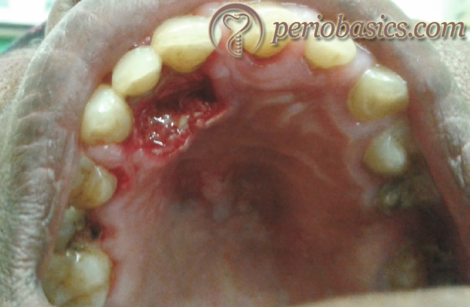
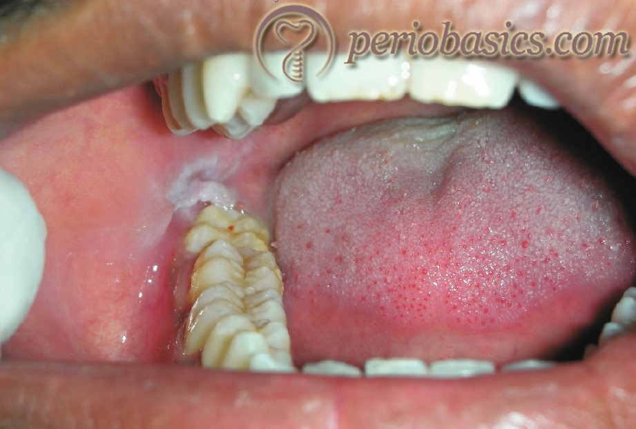
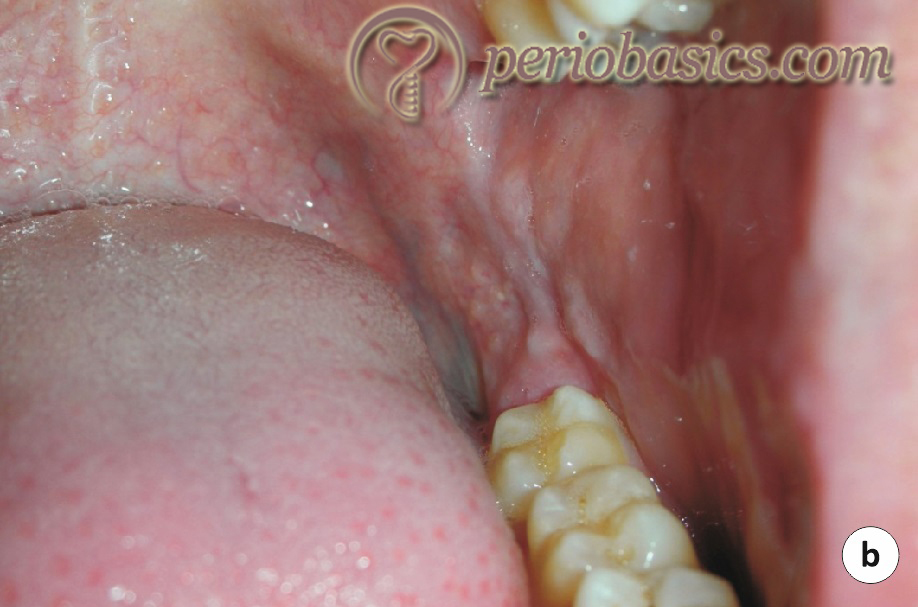
Pericoronal abscess (pericoronitis)
Pericoronitis is the inflammation of soft tissue associated with the crown of a partially erupted tooth. The word pericoronitis is made up of three parts: peri (around), corona (crown) and itis (inflammation). It is most commonly seen in association with partially erupted mandibular third molars. The soft tissue that covers the crown is referred to as an operculum, from which its other name “operculitis” is derived. The condition is common in ……… Contents available in the book ……… Contents available in the book ……… Contents available in the book ……… Contents available in the book……..
Classification:
Pericoronitis may be acute or chronic 57. Acute pericoronitis has a rapid onset and is associated with clinical symptoms such as inflammation and pain. Along with this, the patient may also have some degree of systemic involvement. Acute pericoronitis is commonly seen in patients with poor oral hygiene. The patient usually complains of severe pain which is often referred to the adjacent areas. On clinical examination, the pericoronal area may show swelling and pus discharge. Varying degree of trismus is present and the patient complaints of difficulty in eating and swallowing. Mild to moderate pyrexia may be present in some cases. Regional lymphadenopathy may also be present and sometimes infection may spread to the adjacent spaces.
The chronic or recurrent pericoronitis cases are those in which repeated episodes of acute pericoronitis occur periodically. It is commonly seen in patients with moderate to good oral hygiene. The signs and symptoms are less severe than acute pericoronitis. The patient usually complains of mild to moderate pain which usually subsides within a day or two. Recurrence of the signs and symptoms may occur after many months. Rarely, bilateral pericoronitis may be seen which is strongly suggestive of underlying infectious mononucleosis 56.
Diagnosis:
The diagnosis of pericoronitis can be easily made by taking a careful history of the patient and clinical examination. The inflamed pericoronal flap with or without pus discharge, varying degree of trismus, moderate to severe pain and signs and symptoms of systemic involvement are suggestive of this condition.
Management:
The management of pericoronitis depends on the degree of local and systemic involvement. The case can be managed as follows,
In patients with only localized pain without any regional or systemic involvement, only local measures are suggested. The area should be debrided of plaque and food deposits and if pus is present it should be drained. The area is then irrigated with normal saline, chlorhexidine or hydrogen peroxide. The pericoronal flap should be gently elevated with a curette or scaler and the undersurface of the flap is cleaned with the help of a swab dipped in an antiseptic agent. If the occlusal adjustment is required to prevent trauma to the soft tissue, it should be done.
If the pus discharge is present, an anteroposterior incision is placed with the help of #15 or #12 blade and pus is drained. Pus drainage can also be established using a bent needle electrode of electrocautery unit. In the past, chemical agents were used to cauterize the soft tissue. These chemicals included ……… Contents available in the book ……… Contents available in the book ……… Contents available in the book ……… Contents available in the book……..
In severe cases, where systemic symptoms are also present, aggressive use of antibiotics and analgesics is recommended. The microbiota involved in pericoronal infection consists of a complex mixture of Gram-positive and Gram-negative microorganisms which makes the choice of antibiotics difficult. Use of broad-spectrum antibiotics or a combination of antibiotics has been recommended according to the extent of involvement. Most commonly, a combination of amoxicillin (500mg three times a day for 5 days) and metronidazole (400 mg three times a day for 5 days) is given to control the infection. If the infection does not responds, amoxicillin+clavulanic acid 625 mg two times a day for five days in combination with metronidazole is used. The patients who are allergic to penicillin, erythromycin 500 mg four times a day for five days is used.
After the acute symptoms subside, the decision to go for extraction or operculectomy is made. If the involved tooth is not in an appropriate functional position or is partially erupted or is angulated, its extraction is indicated. On the other hand, if the tooth is in good functional condition and can be retained, operculectomy is planned. Operculectomy can be done by using periodontal knives, scalpel, electrosurgery or laser. A loop electrode can be used to remove the operculum. While removing the tissue it should be remembered that the tissue has to be removed both from the occlusal surface and the distal surface of the tooth (Figure 27.4). If the tissue is ……… Contents available in the book ……… Contents available in the book ……… Contents available in the book ……… Contents available in the book……..

Combined periodontal-endodontic lesions
The combined endo-perio lesions have inflammatory involvement of both endodontic and periodontal tissues to varying degrees. But mostly, when root canal is involved, the patient complaints of moderate to severe pain which requires immediate attention. The simultaneous involvement of pulpal tissue and periodontal tissue can complicate the diagnosis and treatment planning. A detailed description of endo-perio lesions and their treatment has been given in “Periodontic-endodontic interrelationship”.
Other acute periodontal conditions
There are many other conditions which may have their initial clinical manifestation as an acute periodontal lesion, as a part of their complex clinical picture. For example, various mucocutaneous disorders have oral lesions as a part of their clinical manifestations. The vesiculobullous lesions are painful and require immediate attention. An appropriate diagnosis of the condition is required before any treatment is started. More details regarding mucocutaneous lesions are available in “Desquamative gingivitis”.
Various viral infections result in the formation of oral ulcerative lesions which cause a significant discomfort to the infected individual. The most common viral infections associated with the oral cavity are caused by herpesviridae family. HSV-1 and HSV-2 infections have already been discussed. The Varicella-zoster virus causes Varicella which is characterized by the formation of vesicles on the skin after an incubation period of 1-3 weeks. The oral manifestations of the infection include vesicle formation in oral mucosa and gingiva. After resolution of the disease, virus takes shelter in sensory ganglia where it remains in a latent phase for years. The re-activation of this virus causes herpes zoster, which occurs in ……… Contents available in the book ……… Contents available in the book ……… Contents available in the book ……… Contents available in the book……..
Bacterial infections caused by Staphylococci and Streptococci lead to the formation of oral lesions with non-specific appearance. S. aureus causes bullous lesions in the oral cavity along with generalized dermatitis, vesicle formation and desquamation of the mucosal surfaces 63. Group B streptococci infection results in oropharyngeal infection. The systemic manifestation of the infection includes fever and malaise 64-66. Appropriate systemic antibiotic therapy is required to treat these bacterial infections.
Fungal infections may also cause acute oral lesions. Various fungal species are a part of normal flora of the oral cavity. The overgrowth of these species is facilitated by compromised immune response primarily in HIV-positive patients, patients on immunosuppressives (with organ transplant), during infancy and in the elderly. The most common fungal infection is candidiasis 67-70. The most common candida species involved is C. albicans. Although, localized fungal infections are not usually severe, but severe infection may result in systemic involvement. The primary treatment for the disease is to identify the underlying cause and its treatment. The treatments of fungal diseases require anti-fungal agents. Amphotericin B, miconazole or nystatin are used in the form of solutions or gels. In severely immunocompromised patients the drug of choice is fluconazole.
Allergic reactions may also cause acute oral lesions. An allergic reaction may precipitate by a wide variety of substances such as food products, medicaments, metals, oral hygiene products, dental materials, etc. The oral manifestations of the allergic reaction may vary from urticaria to angioedema. However, allergic reactions do not normally affect gingiva 71, 72. The identification of allergen is required for treating allergic reactions.
Another cause of acute periodontal lesions includes vesiculobullous lesions caused due to mucocutaneous disorders. These disorders are manifested in the gingival tissues as desquamative gingivitis. The lesions are characterized by the presence of epithelial desquamation and erythematous, vesicles or bullous or erosive lesions in the gingiva. Although it is normally a part of a chronic process, painful acute phases can also occur, with intense and diffuse redness in the attached and free gingiva. The epithelium is fragile and easily detached, leaving a reddish surface, which bleeds after minimum stimulus. Women in the 4-5th decades of life are more prone to present these lesions
Mechanical or chemical injuries may also cause acute periodontal lesions. Mechanical injuries may occur due to traumatic accidents. These may also occur due to improper brushing or parafunctional habits. Continuous mechanical trauma for a long duration of time causes frictional lesions which are hyperkeratotic in nature. Traumatic injuries may cause tooth fracture, fracturing of the orthodontic or prosthodontic appliance which may result in the formation of an ulcer or periodontal abscess. Chemical burns may also lead to acute gingival lesions. Identification of the cause of acute lesion is required and the treatment is rendered accordingly.
Conclusion
Acute gingival and periodontal lesions require immediate treatment so that the discomfort of the patient is eliminated. The patient should be carefully examined for the clinical signs and symptoms. A prompt and correct diagnosis is the primary requirement for adequately treating a patient with these lesions. Once the acute phase of the disease is controlled, the treatment of the underlying cause of the disease is treated. From the above discussion, it is clear that many diseases can have non-specific oral lesions. Thus, it becomes important to find out the underlying cause for the lesions before any treatment is started. The patient should be kept under recall examination and if any further treatment needs to be done, it should be done. Usually, these lesions heal well without causing much esthetic problems. However, in cases like ANUG, esthetic corrections may be required.
References
References are available in the hard-copy of the website.
Periobasics: A Textbook of Periodontics and Implantology
The book is usually delivered within one week anywhere in India and within three weeks anywhere throughout the world.